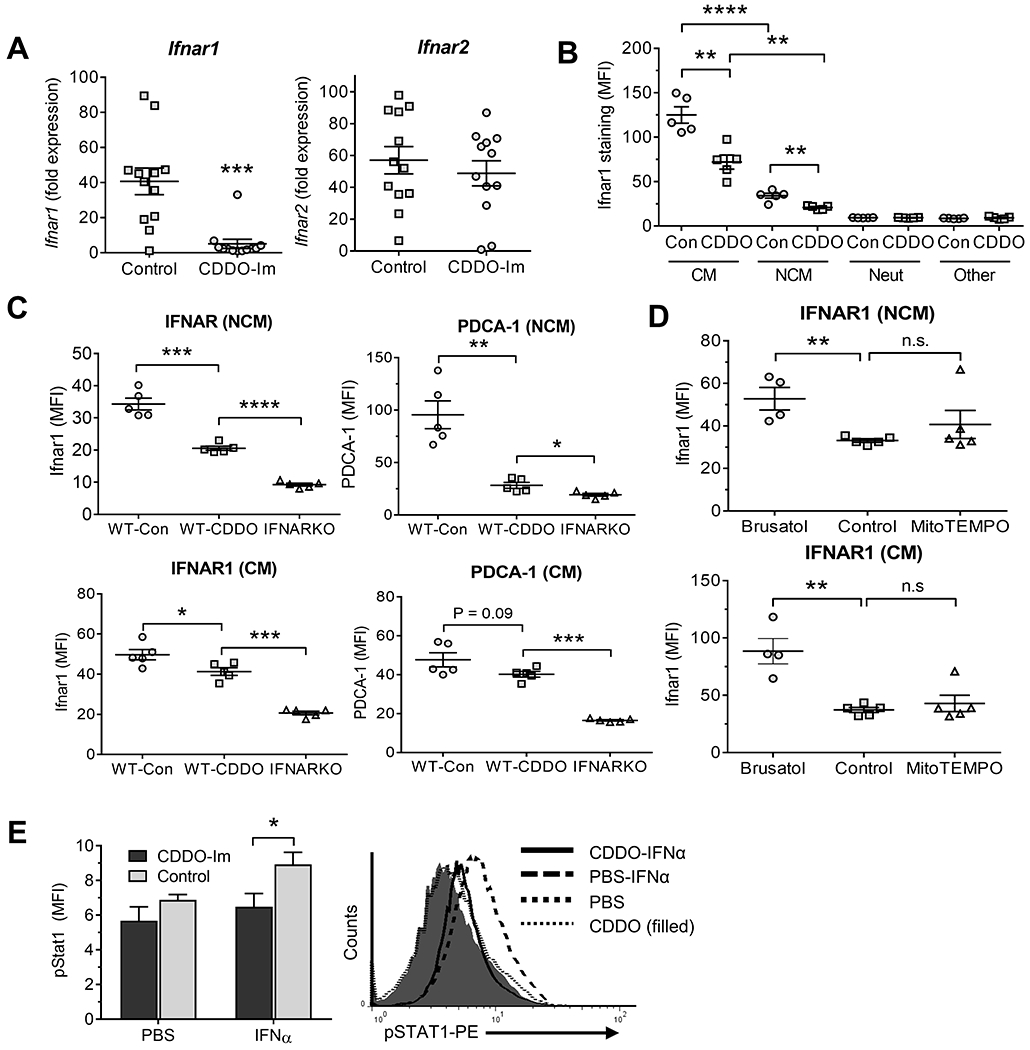
Figure 5. High Nrf2 activity is associated with low Ifnar1 expression.

Pristane-treated B6 mice received either CDDO-Im or vehicle (Control) as in Fig. 2. PEC were analyzed at 6-d. A, Expression of Ifnar1 and Ifnar2 mRNA relative to 18S RNA in total PEC (qPCR). B, Flow cytometry of Ifnar1 expression in PEC gated on CD11b+Ly6G−CD138+ cells (NCM), CD11b+Ly6G−Ly6Chi cells (CM), CD11b+Ly6G+CD138− neutrophils (Neut), and CD11b−Ly6G−CD138− (Other) cells, mainly lymphocytes. C, wild-type B6 (WT) and B6-Ifnar knockout (IFNARKO) mice injected with pristane received CDDO-Im or vehicle (Control, Con) as above. PEC gated on NCM (top) and CM (bottom) were analyzed by flow cytometry at 8-d for IFNAR1 (left) and the IFN-regulated protein PDCA-1 (right). D, Pristane-treated B6 mice received brusatol, MitoTEMPO, or vehicle (Control) as in Fig. 3 for 9-d. IFNAR1 surface staining of peritoneal NCM (Top) and CM (Bottom) was determined. E, pristane-treated B6 mice received CDDO-Im or PBS for 8-d. PEC were treated in vitro with IFNα15 (200 ng/ml) or PBS for 15-min and phosphorylated Stat1 (pStat1) was measured in CD11b+Ly6G− cells by flow cytometry (5/group). Right, representative histograms of IFNAR surface staining in mice treated with IFNα15 vs. PBS from mice treated with pristane + CDDO-Im or pristane alone. * P < 0.05, ** P < 0.01, *** P < 0.001, **** P < 0.0001 (Student t-test). n.s., not significant. (MFI, mean fluorescence intensity).
