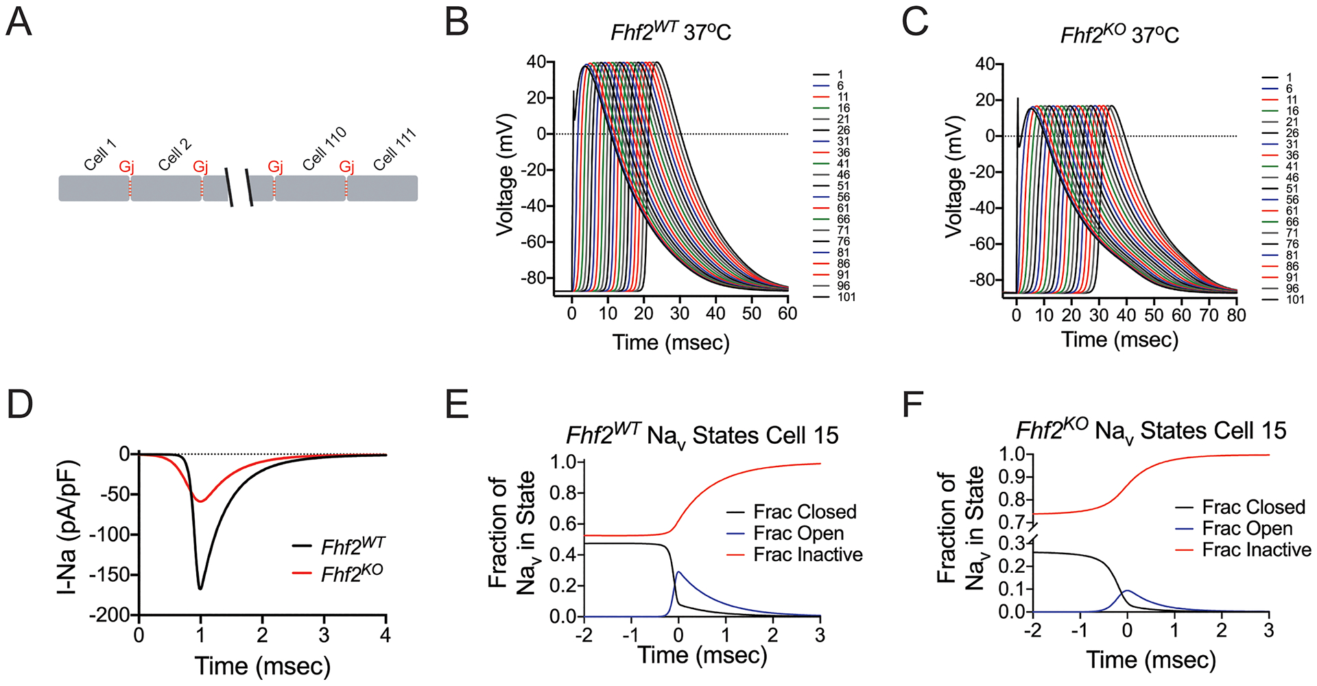
Figure 4. Computational models of Fhf2WT and Fhf2KO cardiomyocyte strands.

(A) Strand model designed to include 111 cells, each measuring 100 microns aligned end-to-end, connected by gap junctions (Gj = 772.8 nS at 37°C). (B) Simulation of action potential (AP) propagation through strand. 0.5msec current injection into the first cell results in AP propagation through entire Fhf2WT strand. The AP propagates with conduction velocity (CV) = 0.475 mm/msec. (C) Similar injection of current into the Fhf2KO strand results in slowed AP propagation (CV = 0.339 mm/msec) with decreased AP amplitude in all cells. (D) Sodium current influx in cell 50 as a function of time. Sodium current influx is reduced in the Fhf2KO strand, despite the same density of sodium conductance (gNaV). (E) In cell 15 of the Fhf2WT strand, sodium channel availability is approximately 50% at resting potential with peak NaV activation peaking much earlier than NaV inactivation. (F) In cell 15 of the Fhf2KO strand, reduced sodium current is the consequence of approximately 74% NaV steady-state inactivation at the resting potential, further NaV closed state inactivation during the voltage rise preceding peak NaV activation, and faster decay of the current due to faster NaV open state inactivation.
