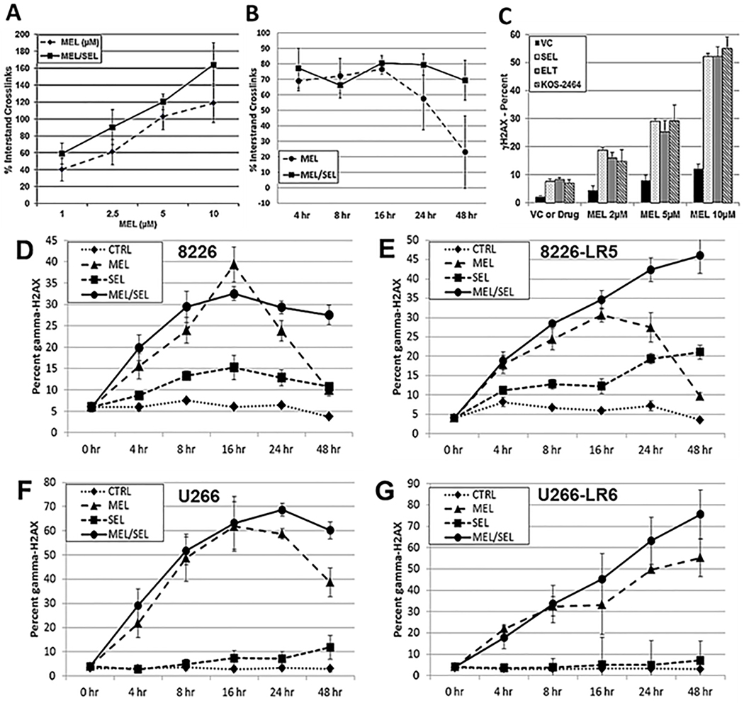
Fig. 4. SEL increases DNA damage and prevents DNA repair.
(A) Alkaline comet assay experiments were used to assay MEL-induced DNA ICL formation. H929 human myeloma cells were exposed to SEL (100 nM) for 20 hours followed by MEL (1, 2.5, 5, and 10 μM) for 2 hours or they were treated with doses of MEL alone at the same increments. Drugs were removed and cells were incubated for 3 hours to allow for ICL formation. Cells were exposed to 900 rads (X-Rad 160 X-ray biological irradiator), and the alkaline comet assay was performed immediately (Trevigen Inc). Image and data analyses were performed by using the Loats Comet Analysis System (Loats Associates) (n=3). (B) Alkaline comet assay outcomes over a 0- to 48-hour time course. H929 human MM cells were treated with MEL (50 μM) for 2 hours to produce DNA damage. MEL was removed and either fresh media or media including SEL (100 nM) were added. Cells were incubated and aliquots were taken at 4, 8, 16, 24, and 48 hours and ICL measured (n=5). (C) Human H929 MM cells were incubated for 20 hours with varying concentrations of MEL (2, 5, and 10 μM) ± SEL (300 nM), ELT (300 nM), or KOS-2464 (100 nM) and then analyzed by FACS for γH2AX (n=3). (D/E) Human 8226 and MEL-resistant 8226-LR5, and (F/G) U266 and MEL-resistant U266-LR6 MM cells were treated for 2 hours with 50 μM MEL, washed, and then incubated with SEL (300 nM) for 4, 8, 16, 24, and 48 hours. Cells were analyzed by FACS for γ-H2AX (n=3).

