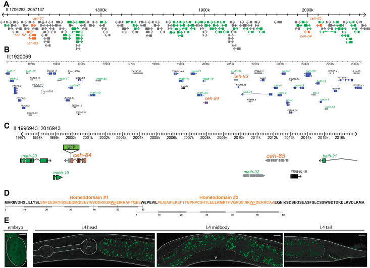
Figure 1.
Location, features and expression of the ceh-84 homeobox gene.
A: Parts of the previously described C. elegans-specific gene cluster on chromosome II are shown. The homeobox genes ceh-81 through ceh-85 are outlined and labelled in orange and are surrounded by MATH and BTB type genes in green. All other genes and non-coding RNAs are colored gray.
B: ceh-84 and ceh-85 genomic region with numerous pseudogenes highlighted in gray, protein coding genes in blue, and non-coding RNAs in black. MATH and BTB type genes labelled in green, homeobox genes labelled in orange, all others in black.
C: Further zoom in on ceh-84 and ceh-85 loci showing schematic of ceh-84 reporter with insertion of GFP sequence before the stop codon. Pseudogenes are colored in gray, protein coding genes belonging to the homeobox family in orange, protein coding genes belonging to the MATH and BTB type in green, all others in black.
D: CEH-84 contains two unusual, short homeodomains. Orange indicates the sequence match to the SSF46689 superfamily domain, a homeodomain signature automatically generated by http://supfam.org. The numbering below indicates the position of canonical, usually 60 aa long homeodomains and their predicted alpha helices (gray bars), with helix 3 binding in the major groove of DNA (Bürglin and Affolter, 2016). This numbering is anchored with the highly conserved WF residues (underlined).
E: Images of CEH-84 protein fusion reporter, PHX2811 (allele syb2811), generated by insertion of gfp sequence by CRISPR/Cas9. On the left, embryonic GFP expression looks diffuse and non-nuclear. On the right, no detectable GFP in L4 hermaphrodites. Solid white lines outline the worm structures, dashed lines highlight the intestinal GFP autofluorescence, and the white V marks the vulva. Using a ~2kb promoter fusion, postembryonic intestinal expression was observed by McKay et al., 2003 and weak embryonic expression was noted by Hench at al., 2015.

