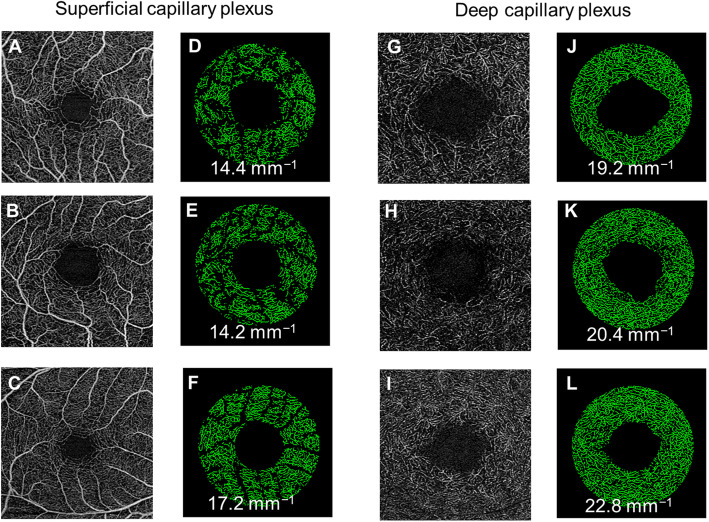
Fig. 3.
Optical coherence tomography angiography (OCTA) images of the superficial (a–c) and deep (g–i) capillary plexuses were extracted from the OCTA machines. d–f, j–l Vessel density maps of the macular annulus region showing retinal microvasculature of participants with Alzheimer’s disease (AD; d, j), mild cognitive impairment (MCI; e, k), and controls (f, l). AD participants showed a decrease in vessel densities in both plexuses compared to controls. MCI participants showed a decrease vessel density only in superficial capillary plexus and not the deep capillary plexus

