Fig. 6.
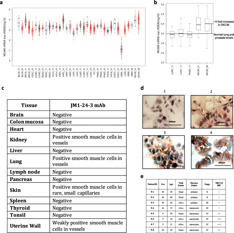
The expression levels of MUC18 in cancers have clinical significance. a The copy numbers of MUC18 mRNA were analyzed across a variety of cancers (red color) and normal tissues (black color) in TCGA cohort studies. The MUC18 gene expression was elevated in melanoma (SKCM) and renal cell carcinoma (KIRC). b The MUC18 gene expression level in melanoma (SKCM) was five times as much as that in normal lung and prostate tissues. c IHC of normal tissue microarray with JM1-24-3, showed positive staining on smooth muscle cells in small vessels of kidney, lung and skin, but not on vessels larger than small capillaries, while other normal tissues were negative. d MUC18 IHC images with JM1-24-3 showing variable staining intensity from negative (upper left panel) to strong positive (lower right panel) on melanoma patient tissue slides. e Staining intensity correlation of eight melanoma patients showed that metastatic melanoma patients had higher intensity staining of MUC18 with JM1-24-3 mAb. Also, all metastatic patients (5/5) showed stronger intensity

