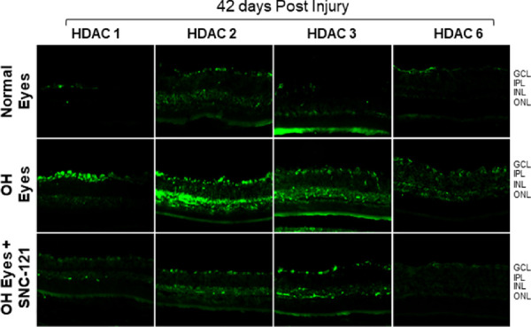
Figure 10.

Immunohistochemistry of HDAC 1, 2, 3, and 6 in the retina at day 42 post-injury. Animals were euthanized and eyes were enucleated at day 42 post hypertonic saline injections. Cryosections were immunostained by a selective anti-HDAC 1, anti-HDAC 2, anti-HDAC 3, or anti-HDAC 6 antibodies. There was no positive staining when primary antibodies were omitted (not shown). Data are a representation of at least six independent experiments. Comparable immunostaining for each HDAC was seen in the retina of other animals (magnification times 20). GCL, ganglion cells layer; IPL, inner plexiform layer; INL, inner nuclear layer; ONL, outer nuclear layer.
