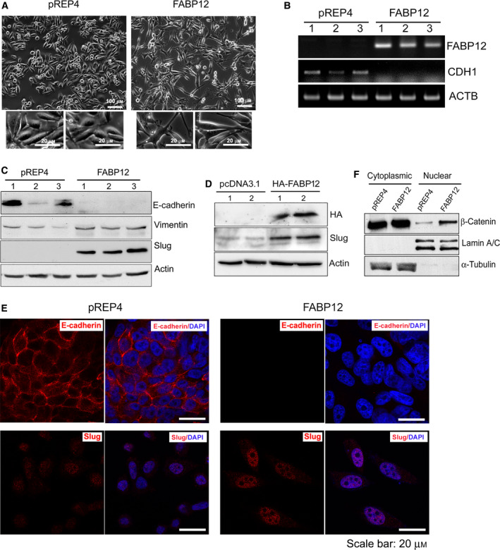
Fig. 3.

FABP12 induces EMT. (A) Stable ectopic expression of FABP12 induces morphological changes in PC3 cells. FABP12‐expressing cells show an enlarged and fibroblast‐like appearance with elongated processes (right panel, with magnified images from two different regions shown below) compared to the control cells which have a slightly elongated shape (left panel, with magnified images from two different regions shown below) compared to the control cells which have a slightly elongated shape (left panel). (B) Semiquantitative RT‐PCR showing the loss of CDH1 (encoding E‐cadherin) transcripts in PC3‐pRE4‐FABP12 cells. (C) Western blotting showing the induction of Slug, loss of E‐cadherin, and increased levels of vimentin in PC3‐pREP4‐FABP12 compared to PC3‐pREP4 control cells. (D) Western blotting showing the induction of Slug in PC3 cells transiently transfected with pcDNA3.1‐HA‐FABP12 compared to control cells. (E) Immunofluorescence staining of E‐cadherin and Slug in PC3‐pREP4 control (left panel) and PC3‐pREP4‐FABP12 cells (right panel). (F) Western blotting showing increased levels of β‐catenin in the nucleus of PC3‐pREP4‐FABP12 cells compared to PC3‐pREP4 cells. Human β‐actin, lamin A/C, and α‐tubulin served as loading controls for whole‐cell, nuclear, and cytoplasmic lysates, respectively. N = 3.
