Fig. 7.

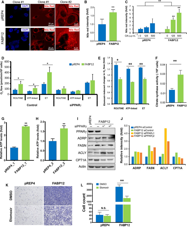
FABP12 expression enhances cellular lipid accumulation and mitochondrial fatty acid β‐oxidation. (A) Confocal microscopy images of Nile Red staining of lipid droplets in PC3 control cells—clone #1 (pREP4, upper panel) and PC3 cells stably expressing FABP12—clone #1 (lower panel). The panels on the right show magnified images of PC3‐pREP4—clone #2 and PC3‐pREP4‐FABP12 clone #2. DAPI was used as a nuclear stain for PC3—clones #1 (left panels). (B) Quantification of Nile Red staining of lipid droplets in stable PC3 control and FABP12‐expressing cells. (C) Lipid droplet accumulation in stable PC3 control and FABP12‐expressing cells cultured in medium supplemented with oleic acid. Two‐way ANOVA was used for statistical analysis. (D) Oxygen consumption by mitochondria of stable PC3 control and FABP12‐expressing cells. Significantly increased ROUTINE, ATP‐linked, and ET (maximum) O2 consumption was observed in FABP12‐expressing cells. This increase in O2 consumption was no longer observed upon PPARγ depletion. (E) O2 consumption in ROUTINE, ATP‐linked, and ET states was all decreased in PC3 cells stably transfected with pREP4‐FABP12 (FABP12) compared to control cells (pREP4) upon treatment with 100 μm etomoxir, a CPT1 inhibitor. (F) Ectopic expression of FABP12 induces significantly higher activity of citrate synthase, the rate‐limiting enzyme for the Kreb's cycle. (G, H) Increased ATP levels were observed in two clonal populations of stably transfected PC3‐pREP4‐FABP12 cells compared to control PC3‐pREP4 cells. (I, J) Effect of FABP12 ectopic expression and PPARγ depletion on expression of key enzymes related to lipid metabolism. (K, L) Representative images (K) and results (L) showing the effects of FABP12 expression and etomoxir treatment on cell migration using the Transwell assay. Statistical analyses in B, D, E, F, G, H, J, and L were carried out using Student's t‐test. *P < 0.05; **P < 0.01. Error bar: SD.
