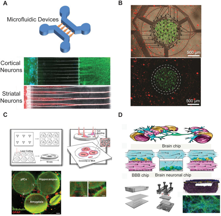
Figure 2.
In vitro culturing of multiple brain regions. (A) Representative image of a microfluidic device (top) and immunofluorescence micrographs (bottom) of cortical (in green) and striatal (in red) neurons growing inside the chips [modified from Peyrin et al. (2011) with permission from The Royal Society of Chemistry]. (B) Novel multielectrode array device used for co-culturing primary rodent hippocampal and cortical neurons [modified from Soscia et al. (2017) with permission]. (C) Schematic representation of a novel brain-on-a-chip model comprising the three different brain regions, prefrontal cortex, hippocampus and amygdala, shown via confocal images (bottom), stained for β-tubulin III (in green), and GFAP (in red), [modified from Dauth et al. (2017) with permission]. (D) Schematic representation of a microfluidic device allowing to metabolically couple neuronal and endothelial cells [modified from Maoz et al. (2018) with permission].

