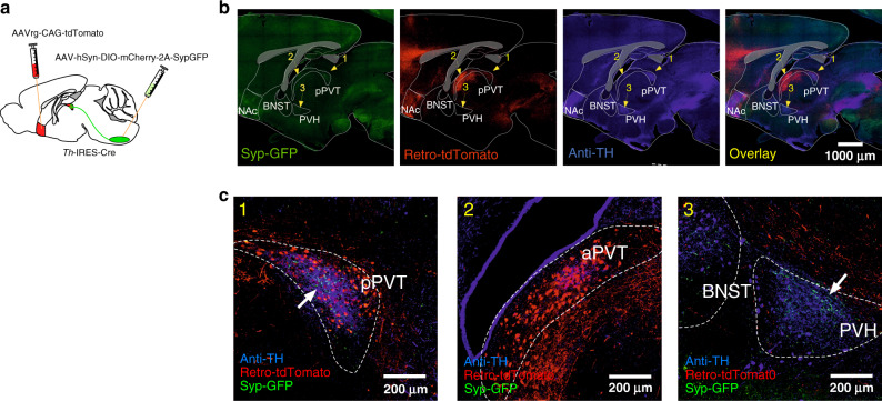
Fig. 1. Axonal projections from VLMCA neurons innervate the pPVT.
a Schematic of virus injections strategy used to examine putative forebrain areas that had robust axonal inputs of VLMCA neurons and that project to the NAc. b Representative images of a sagittal brain slice showing the distribution of VLMCA projections, NAc-projecting neurons (Retro-tdTomato) and TH immunoreactivity (anti-TH) across various forebrain regions. c Representative images showing magnifications of the areas depicted in b, namely the pPVT (1), the aPVT (2), and the BNST/PVH region (3). White arrows depict regions of dense Syp-GFP expression.

