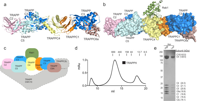
Fig. 1. Structure models for TRAPP core with and without Rab1 and purification of TRAPPII complex.
a Model of the mammalian TRAPP conserved subunits. The structural model was generated through a combination of the following structures (PDB: 2J3T, 2J3W, and 3CUE)37,38,78. Phyre2 was utilized to generate structures of mammalian homologs for TRAPP subunits with no solved crystal structure79. The location of TRAPPC2L bound to the conserved subunits is unknown. b Surface representation of a model of the human TRAPP core subunits bound to Rab1(PDB:3CUE)38. c Cartoon model for the human TRAPPII complex. The core subunits are colored according to the models in (a) and (b). The gray boxes represent TRAPPII specific subunits with an unknown orientation. d Size exclusion chromatography (SEC) trace of TRAPPII on a Superose 6 gel filtration column with molecular weight markers indicated (kDa). The y-axis is normalized to max mAU. e SDS-PAGE gel of purified TRAPPII complex from the gel filtration peak (~13.5 ml) shown in panel (d). High and low labels refer to protein amount loaded (high = 4 μg, low = 0.67 μg).

