
Fig. 2.
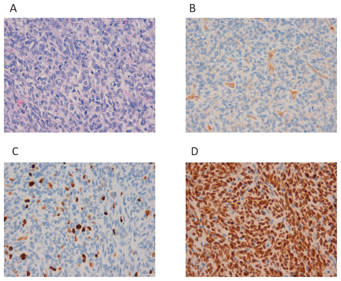
Histopathology of Case 1.
Fig. 2A: Growth of swollen spindle cells with necrosis and mitosis.
Fig. 2B: Partial CD34 positivity is observed.
Fig. 2C: The Ki-67 index is 20%.
Fig. 2D: Strong nuclear staining with anti-STAT6 antibody can be observed.