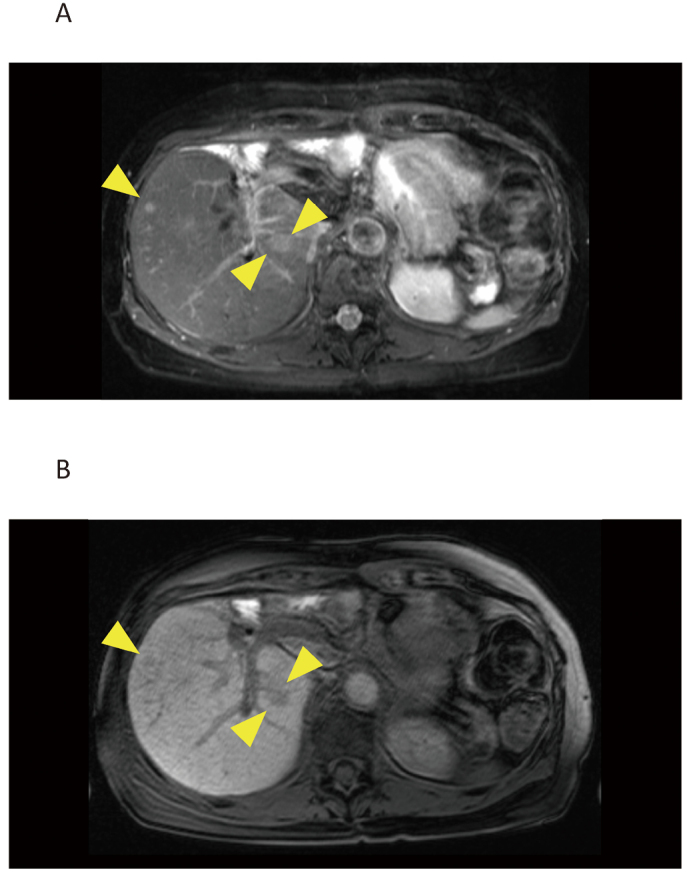
Fig. 2.

MRI of liver metastases in a 74-year-old woman who received surgery for hilar cholangiocarcinoma five years ago
Fig. 2A: A fat suppression T2-weighted image
Fig. 2B: A gadolinium-EOB-DTPA-enhanced T1-weighted image in the hepatocyte phase

MRI of liver metastases in a 74-year-old woman who received surgery for hilar cholangiocarcinoma five years ago
Fig. 2A: A fat suppression T2-weighted image
Fig. 2B: A gadolinium-EOB-DTPA-enhanced T1-weighted image in the hepatocyte phase