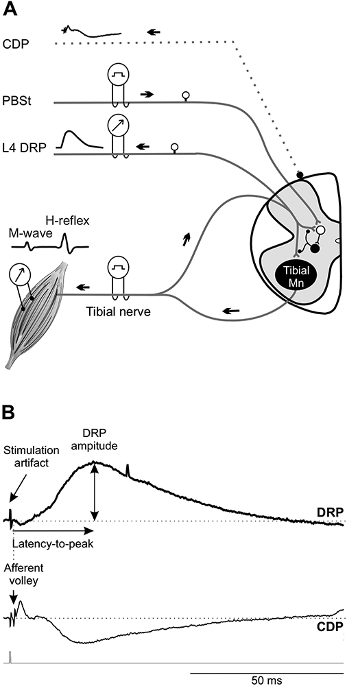
Figure 1. Experimental set-up.

A, The PBSt nerve, tibial nerve and cut dorsal rootlet were mounted on bipolar hook electrodes. The cord dorsum potential (CDP) was recorded using a monopolar silver ball electrode placed at the L4-L5 dorsal root entry to determine the activation threshold for the most excitable afferent fiber. Dorsal root potentials (DRPs) were recorded in response to a stimulation to the tibial or PBSt nerves. H-reflexes were evoked by a stimulation to the tibial nerve and obtained using bipolar wire electrodes inserted under the plantar surface of the foot. In the conditioning protocol, PBSt stimulation was used as the conditioning stimulus and preceded the tibial stimulation. B, Typical DRP recording displaying a long-lasting upward deflection. By convention, DRP negativity is represented upwards and CDP negativity downwards. The latency-to-peak was determined as the time between the onset of the afferent volley (CDP) and the maximal DRP amplitude from baseline to peak.
