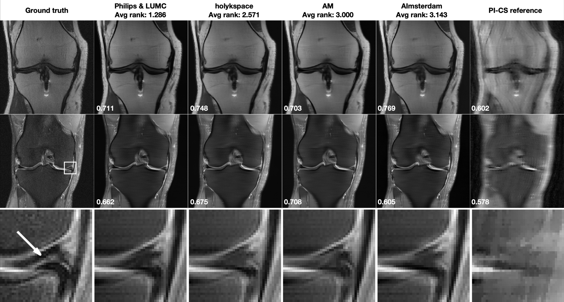
FIGURE 3.

Multi-Coil R=8 track results: Selected results from the top 4 submissions in each track. The submissions are ordered from left to right based on the average of radiologists’ rankings. A combined parallel imaging and compressed sensing reconstruction using Total Generalized Variation (PI-CS) is shown for reference. SSIM to the ground truth for this particular slice is displayed in the bottom-left corner of each image. First row: Results for one slice from an acquisition without fat suppression. This case shows shows moderate artifact from a metal implant. Second row: One slice from an acquisition with fat suppression. This case shows shows a meniscal tear in the ROI indicated by a white rectangle in the ground truth image. Third row: Zoomed view of the ROI that shows a meniscal tear (highlighted by a white arrow in the ground truth reconstruction). This pathology is not well seen in any of the accelerated reconstructions.
