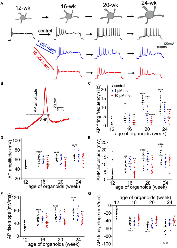
FIGURE 4.
Methadone exposure suppresses the increase in membrane excitability during early neurodevelopment. (A) Representative traces of AP firing in neurons from human cortical organoids at indicated ages (12- to 24-week) in the absence and presence of methadone treatments. APs were evoked from –70 mV to 1,000 ms-long injections of 50 pA current. The exposure of 1 and 10 μM methadone hinders the growth of AP firing during the indicated period of cortical organoids. (B) Representative trace of the 1st AP for the measurement of AP and AHP amplitudes. (C) Summary of the AP firing frequency in response to 1,000 ms current step (50 pA) from –70 mV in neurons at the edge of organoids at indicated ages. (D–G) Summary of the AP amplitude, AP maximal rise slope, AP maximal decay slope, and AHP amplitude evoked by the same current injection (50 pA). In each scatter plot, Black, Blue, and Red dots denote untreated, 1 and 10 μM methadone treated group, respectively. Graphs display mean and SEM. n = 8–25 neurons (6–15 organoids). Two-way ANOVA followed by Tukey’s multiple comparisons test: (C) F(6, 162) = 15.47, p < 0.0001; (D) F(6, 162) = 3.11, p < 0.01; (E) F(6, 162) = 3.67, p < 0.01; (F) F(6, 162) = 2.74, p < 0.05; (G) F(6, 162) = 3.62, p < 0.01. Control groups at 16-week, 20-week, 24-week were compared to 12-week group, ##p < 0.01, ####p < 0.0001. Each methadone-treated groups were compared to the control group at the same age, *p < 0.05, **p < 0.01, ***p < 0.001, and ****p < 0.0001.

