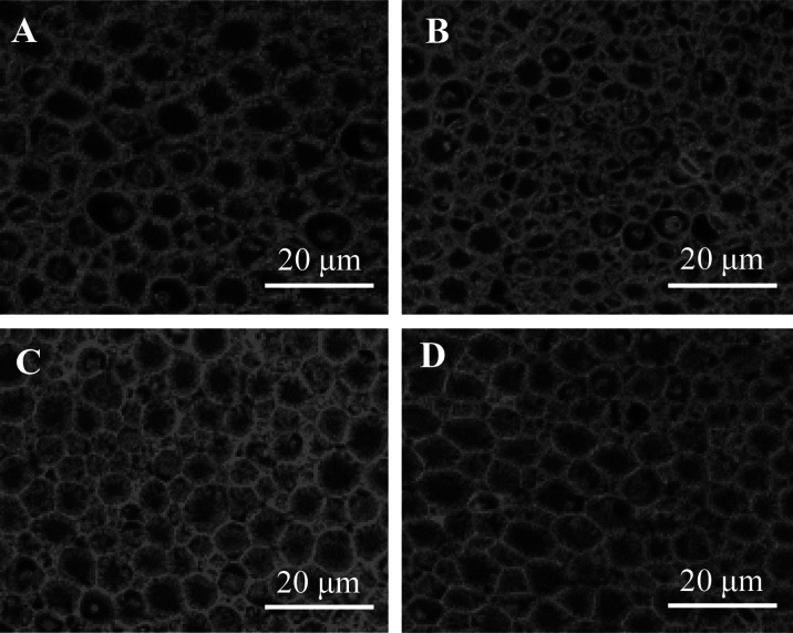
Fig. 2.
High-magnification image of nerve fibers in sciatic nerve in Experiment 1 (Exp.1). Sciatic nerve was collected on day 32 in the Exp.1. A; Glucose + Phosphate buffered saline (PBS) group, B; Oxaliplatin (OXL) + PBS group, C; OXL + recombinant human lactoferrin (rhLF) group, D; OXL + recombinant human lactoferrin Fc fusion (rhLF-Fc) group. The samples were stained with Hematoxylin-Eosin. Scale bar=20 µm. In Image B, atrophy and loss of nerve fibers was observed.

