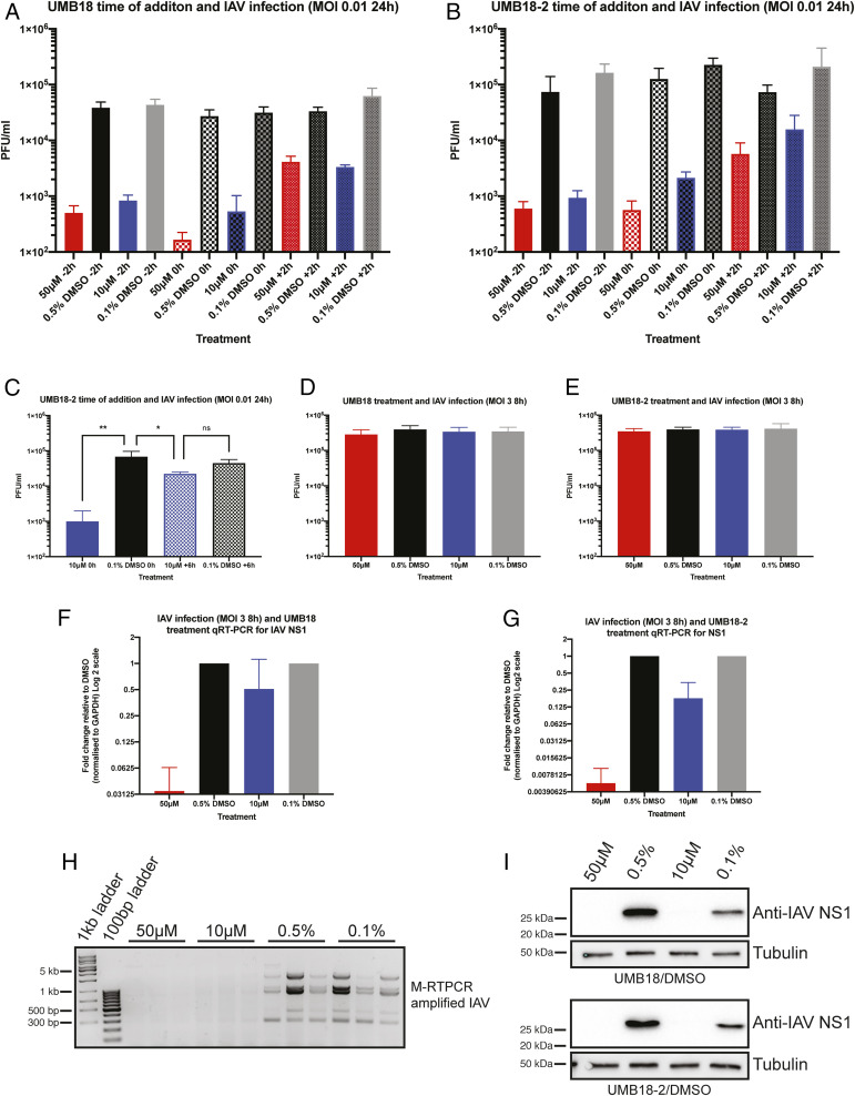
Fig. 6.
SKI targeting lead compounds inhibit viral RNA and protein production. Time-of-addition experiments were performed with IAV infection and UMB18 (A) and UMB18-2 (B). A549 cells were plated and treated with drug 2 h prior to infection (−2 h), at the time of infection (0 h), or 2 h after virus was added to cells (+2 h). After 24 h infection supernatant was collected and titer determined by plaque assay. Mean PFU/mL and SD are displayed from two independent experiments performed in triplicate with error bars being SD. (C) Experimental setup as described in B, but with UMB18-2 or DMSO control added at 0 h or +6 h. Data are from a representative experiment of two performed in triplicate. A one-way ANOVA was performed; ns, nonsignificant, *P < 0.05, **P < 0.01. (D) A549 cells were infected with IAV at MOI 3 for 8 h with UMB18 or (E) UMB18-2 treatment. Supernatant was collected and used to titer by plaque assay. Data are from three independent experiments performed on triplicate wells with mean PFU/mL displayed. (F and G) The same infected cells from D and E were collected in TRIzol and NS1 mRNA transcript analyzed by qRT-PCR. Input levels were normalized to GAPDH and fold change of transcript was determined relative to DMSO control. (H) Using the same extracted RNA as in G, an M-RTPCR protocol was used to amplify all IAV segments which were analysed on an agarose gel. Displayed are the amplifications from two independent wells for UMB18-2 at 50 μM and 10 μM and three wells for DMSO controls. (I) Infected and treated cells were also collected in RIPA lysis buffer and used for Western blotting of NS1. Displayed is a representative blot of the three independent repeats for each compound.

