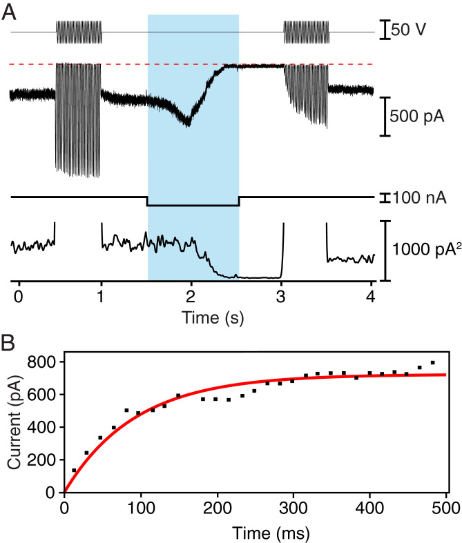
Fig. 1.
Rapid recovery of mechanoelectrical transduction in outer hair cells from the rat’s cochlea. (A) A fluid-jet stimulator was driven at 60 Hz with two sinusoidal stimulus trains (top trace), one before and one after an iontophoretic current pulse (bottom trace) that released EDTA. The pale blue band here and in subsequent figures delineates the period of iontophoresis. The transduction current (second trace) was initially large but fell to nearly zero after iontophoresis before recovering more than one-half of its original magnitude during the second stimulus train. The variance of the transduction current (third trace) fell during iontophoresis as transduction was interrupted, but recovered partially after a second epoch of stimulation. The abscissa represents zero variance. (B) For the record shown in A, the recovery of the transduction current after the iontophoretic pulse followed an exponential relation (red line) with a time constant of 100 ms.

