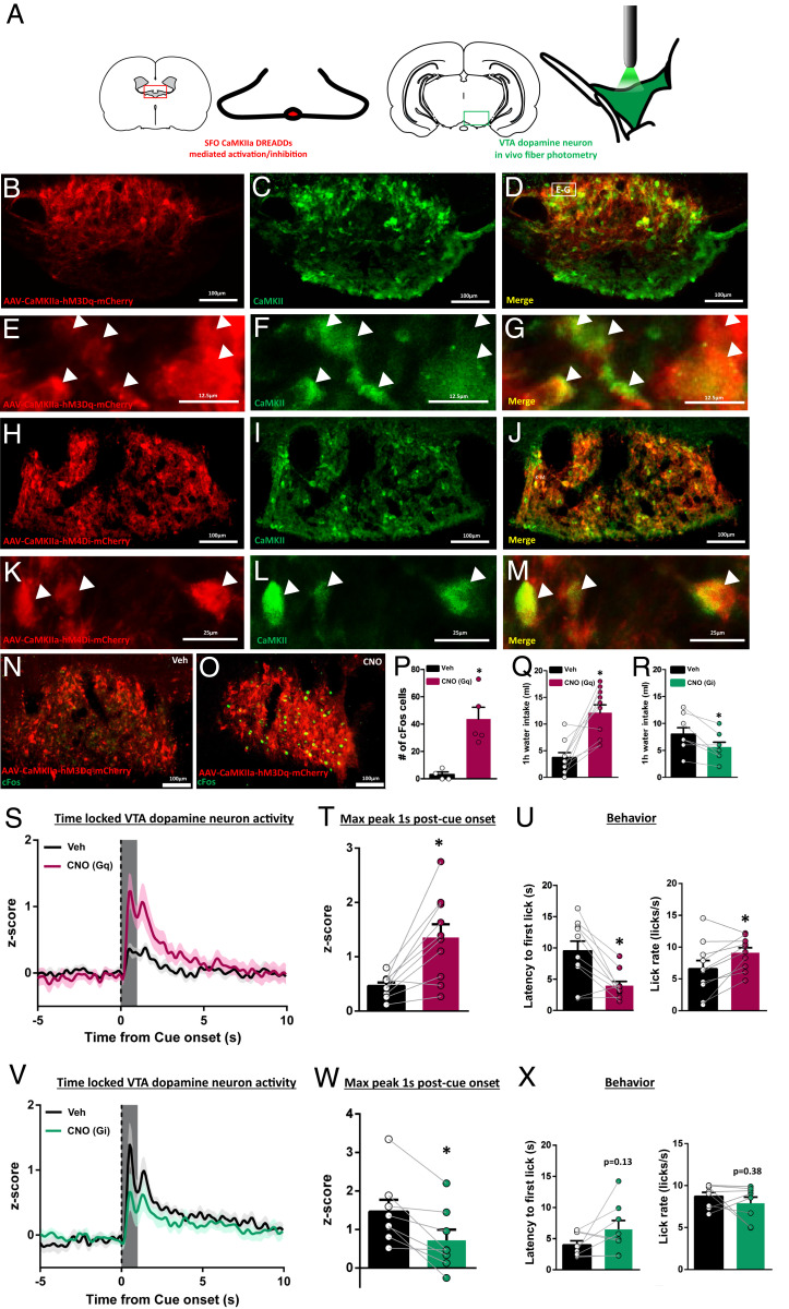
Fig. 4.
SFOCaMKIIa activity is necessary and sufficient for water-cue-evoked VTA dopamine neuron activity. (A) Experimental design using a combination of selective manipulation of SFOCaMKIIa with DREADDs and in vivo fiber photometry from VTA dopamine neurons. (B–M) Representative images of SFO CaMKIIa-hM3Dq-mCherry (B–G) or CaMKIIa-hM4di (H–M) (red) and CaMKII expression (green). High-magnification Insets are shown in E–G and K–M with white arrows showing DREADD and CaMKII colocalization. (N and O) cFos expression (green) following vehicle or CNO (1 µg/μL) in CaMKIIa-hM3Dq transfected SFO neurons and quantification in P. (Q and R) One-hour water intake following vehicle or CNO treatment in (Q) SFO CaMKIIa-hM3Dq (euvolemic) or (R) SFO CaMKIIa-hM4Di (water-restricted) transfected rats. (S) VTA dopamine neuron activity in euvolemic, CaMKIIa-hM3Dq transfected rats following vehicle or CNO treatment (t = 0 for cue onset) with quantification in T. (U) Latency to first lick and lick rate in euvolemic vehicle or euvolemic CNO rats from S and T. (V) VTA dopamine neuron activity in water-restricted, CaMKIIa-hM4Di transfected rats following vehicle or CNO treatment (t = 0 for cue onset) with quantification in W. (X) Latency to first lick and lick rate in water-restricted vehicle or water-restricted CNO rats from V–W. Dark lines in S and V are means and shading are SEM. Bars and whiskers in all graphs are means SEM. Gray boxes in T and W represent 1-s time window postcue onset for quantification and analysis. *P < 0.05 vs. vehicle.

