Fig. 2.
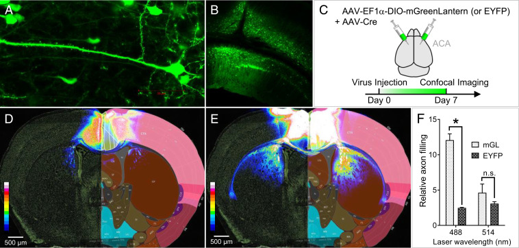
mGreenLantern expresses efficiently in the mouse brain and robustly illuminates long-range neuronal projections. (A) At 7 d post injection (d.p.i.), individual mGreenLantern-expressing neurons of the visual cortex were readily discernible using 63× magnification, (B) as well as the characteristic organization of visual cortex layer VI and hippocampus subfield CA1 at 10× magnification. (C) Injection strategy to label neurons using mGreenLantern and Cre virus mixture in a 10:1 particle ratio. (D) EYFP injected into the ACA as depicted in A did not effectively highlight neuronal projections from ACA to striatum at 14 d.p.i. Rather, fluorescence was primarily restricted to the ACA injection area. Grayscale confocal fluorescence microscopy images were converted to 16-color heat maps and overlaid with corresponding sections from the Allen Brain Atlas from an age-matched mouse for visual reference. (E) mGreenLantern fluorescence at 14 d.p.i. was clearly visible in neurons originating from ACA cell bodies with axonal projections radiating through striatum, corpus callosum, and claustrum. (F) Area of projections in corpus callosum and striatum relative to ACA expression is quantified; n = 3 mice, two-way ANOVA, *P < 0.05.

