Fig. 3.
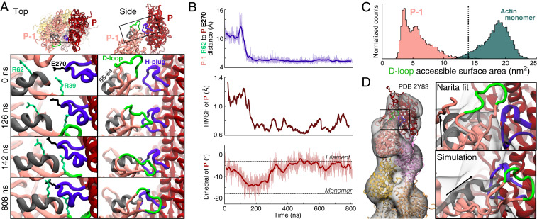
Transition of the ADP pointed end subunit P-1 to a monomeric dihedral angle gives rise to stabilizing contacts with subunit P that flatten P and sequester the D-loop of P-1. (A) Ribbon diagrams from two views of the pointed end of an ADP-actin filament showing unique contacts formed during MD simulations between subdomain 2 of subunit P-1 and subdomains 3 to 4 of subunit P. The four panels in each column show selected time points. (Left) Looking down the filament axis at the pointed end, R39 and R62 of subunit P-1 (foam green) contact the hydrophobic plug (violet) of subunit P (E270 shown in black). These long-lasting interactions stabilize a shift in the position of a helix composed of residues 55 to 64 (gray) of subunit P-1. (Right) A side view depicts the progressive shift of subdomain 2 of P-1 toward subunit P resulting in the D-loop of subunit P-1 (green) forming a stable, lateral contact with subunit P. (B) Time course of the formation of the connection between subunits P and P-1 that stabilizes and flattens the pointed end. Raw data (light) and 20-ns moving averages (dark) are shown. (Top) Distance between the Cα atoms of R62 of subunit P-1 and E270 in the H-plug of subunit P. (Middle) The 40-ns RMSF of subunit P’s center of mass drops in step with the formation of contacts with P-1. (Bottom) The dihedral angle of subunit P transitions toward the monomer conformation, then back to the flattened structure after forming contacts with P-1. (C) Histograms of the accessible surface areas at a radius of 7 Å around the D-loops of subunit P-1 and actin monomers. The dotted line marks the value assuming P-1 takes on the flattened conformation of interior subunits (PDB 6DJO). Data and snapshots are from simulations 7 to 9 for P-1 and simulations 10 to 12 for the actin monomers (SI Appendix, Table S1). (D) Comparison of pointed end models with the 3D reconstruction of the ADP-actin pointed end by Narita et al. (15). (Left) Narita model. (Right) Enlargement of the bridge-like density connecting P-1 and P in the model. Our simulated ADP structure (Bottom) places the structured alpha-helix of subunit P-1 (black arrow) and H-plug of P within the bridge density with the D-loop stably attached to subunit P.

