Fig. 3.
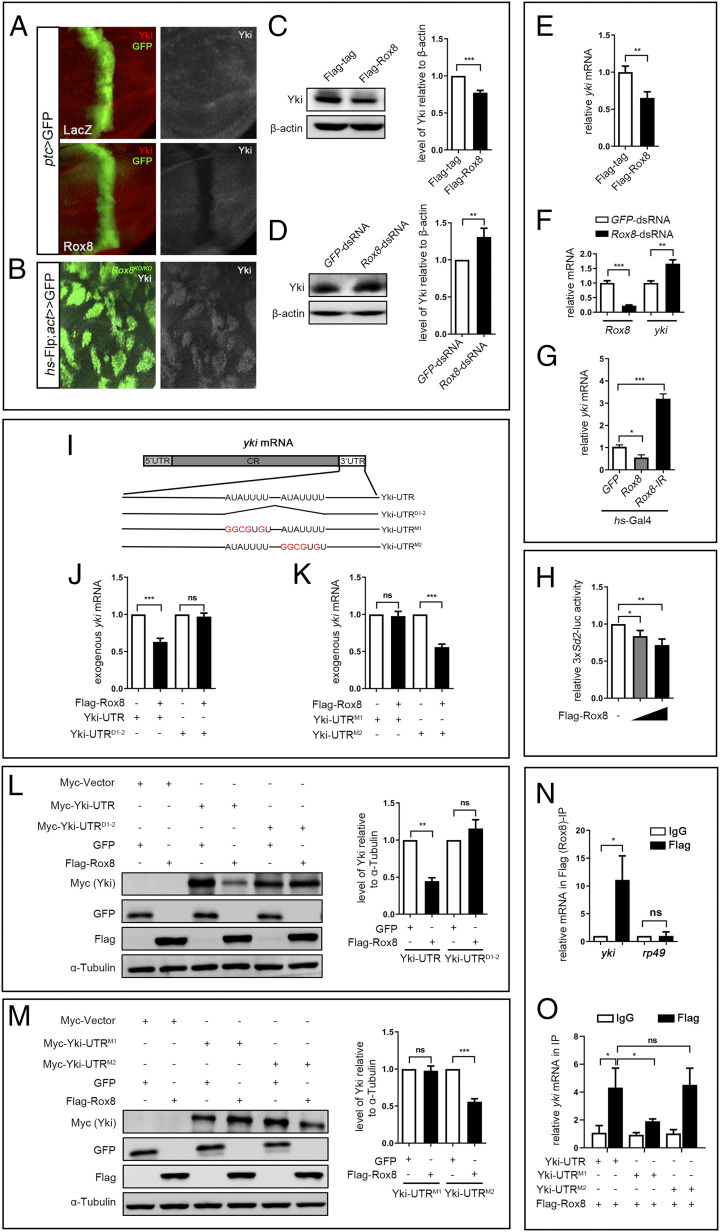
Rox8 accelerates yki mRNA decay. (A and B) Fluorescence micrographs of wing discs are shown. Yki protein level in the wing disk was attenuated by Rox8 overexpression (A), but was up-regulated in Rox8 mutant clones (B). Yki protein level in S2 cells was decreased by Rox8 overexpression (C), but was increased by Rox8 knockdown (D). (E) yki mRNA level was significantly down-regulated by Rox8 overexpression in S2 cells. (F) The level of yki mRNA was up-regulated by knockdown of Rox8 in S2 cells. (G) RT-qPCR data showing yki mRNA level in larval wing discs was reduced or elevated by Rox8 overexpression or depletion driven by hs-Gal4. (H) The expression of 3× Sd2-luc, a reporter for Yki/Sd activity, was inhibited by Rox8 in a dosage-dependent manner. (I) Schematic view of yki mRNA with the 5′ UTR, coding region (CR), and 3′ UTR, which contains two potential Rox8-binding motifs (AUAUUUU). Both motifs are deleted in UTRD1-2, while the first or second motif was mutated in UTRM1 or UTRM2, respectively. (J–M) Yki-UTR, Yki-UTRD1-2, Yki-UTRM1, or Yki-UTRM2 was subcloned into pUAST-Myc-tag vector. To detect exogenous yki mRNA, qPCR primers were designed to span the Myc-tag and Yki coding region. Overexpressing Rox8 resulted in reduction of exogenous Yki-UTR mRNA but not Yki-UTRD1-2 (J). Rox8 overexpression attenuated exogenous Yki-UTRM2 mRNA but not Yki-UTRM1 (K). (L and M) Immunoblot analysis of Myc-tagged-Yki-UTR, UTRD1-2, UTRM1, or UTRM2 expression upon coexpression of Flag-Rox8 in S2 cells. (Left) Immunoblot staining. (Right) Quantification data. As shown in L, expression of Myc-Yki-UTR but not of Myc-Yki-UTRD1-2 was dramatically inhibited by Rox8 overexpression. (M) Expression of Myc-Yki-UTRM2 but not Myc-Yki-UTRM1 was attenuated by overexpression of Rox8. (N and O) RIP assay was performed to detect the physical interaction between Rox8 and yki mRNA. Rox8 specifically bound to endogenous yki mRNA but not to rp49 mRNA that served as a negative control (N). The binding of Rox8 to yki 3′ UTR was blocked by M1 mutation, but not by M2 in S2 cells (O). ***P < 0.001, **P < 0.01, *P < 0.05. ns, no significant difference.

