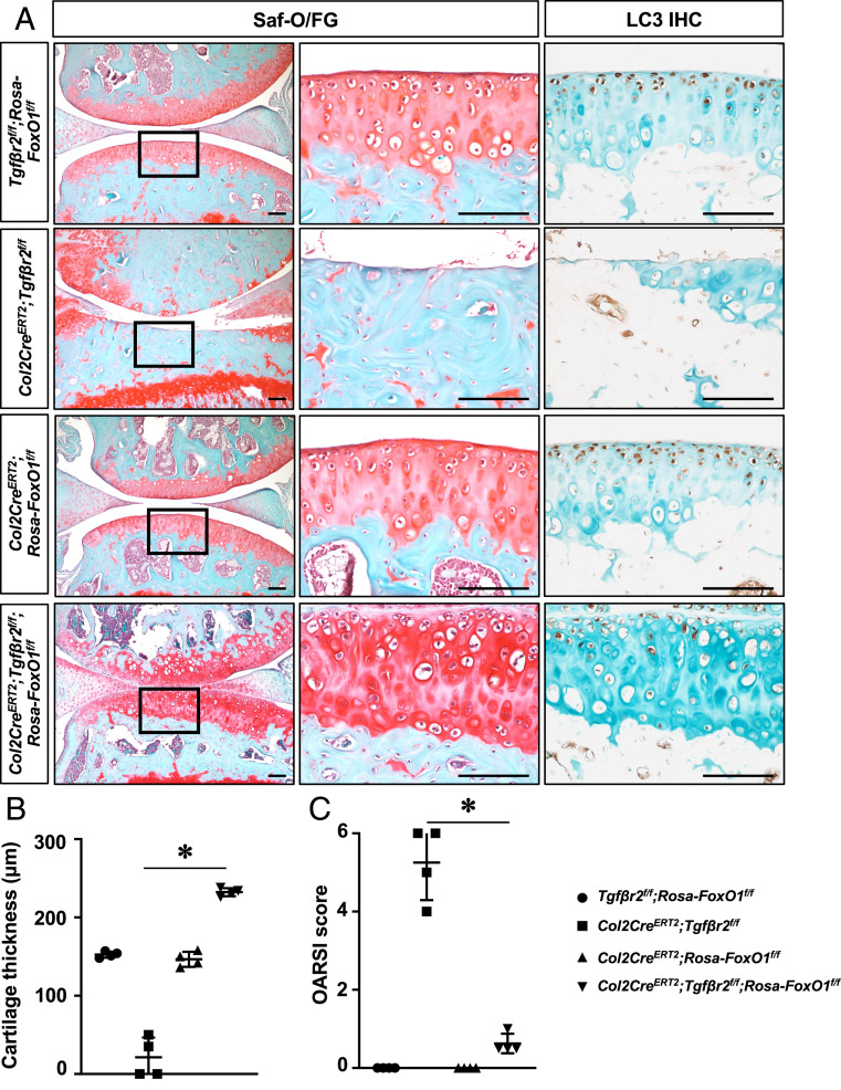
Fig. 6.
FoxO1 overexpression in chondrocytes rescues OA phenotypes induced by loss of TGFβ pathway. (A) Safranin O/fast green staining and immunostaining for LC3 on knee sections of Tgf-βr2f/f;Rosa-FoxO1f/f, Col2CreERT2;Tgf-βr2f/f, Col2CreERT2;Rosa-FoxO1f/f, and Col2CreERT2;Tgf-βr2f/f;Rosa-FoxO1f/f mice at 3 mo of age. All mice, including Cre negative controls, received tamoxifen. n = 4 (Scale bar, 100 μm.) (B) Histomorphometric analyses of cartilage thickness on knee sections of Tgf-βr2f/f;Rosa-FoxO1f/f, Col2CreERT2;Tgf-βr2f/f, Col2CreERT2;Rosa-FoxO1f/f, and Col2CreERT2;Tgf-βr2f/f;Rosa-FoxO1f/f mice at 3 mo of age. All results were compared to Tgf-βr2f/f;Rosa-FoxO1f/f mice and expressed as means ± SD *P < 0.05. n = 4. (C) OARSI scores for the medial tibial plateau and femoral condyle from 3-mo-old mice. *P < 0.05 compared between Col2CreERT2;Tgf-βr2f/f, and Col2CreERT2;Tgf-βr2f/f;Rosa-FoxO1f/f mice. n = 4.

