Fig. 1.
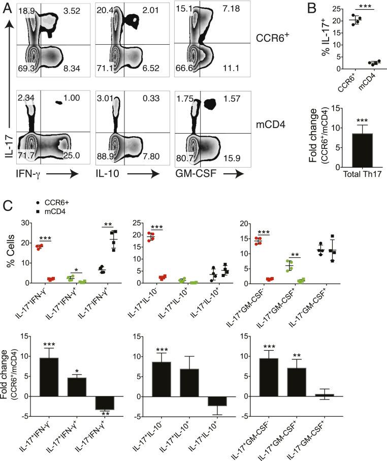
Enriched Th17 and reduced Th1 populations in CD4+CCR6+CXCR3− T cells. CCR6+CXCR3− memory CD4+ T (CCR6+) cells and mCD4 T cells were isolated from the peripheral blood of four healthy donors and stimulated with PMA and ionomycin for 4 h. Production of cytokines in CD4+ T cells were assessed by flow cytometry with intracellular cytokine staining assay. (A) IL-17, IFN-γ, IL-10, and GM-CSF production in CCR6+ and mCD4 cells. Dot plots shown were gated on CD4+ cells from one representative individual. (B) Frequency (Upper) and fold change of the frequency (CCR6+ vs. mCD4) (Lower) of total Th17 cells in CCR6+ and mCD4 cells. (C) Frequency (Upper) and fold change of the frequency (Lower) on various cytokine-secreting Th-subsets. *P < 0.05, **P < 0.005, ***P < 0.0005, two-tailed, paired Student’s t test on each cell subset between CCR6+ and mCD4 cells (individuals n = 4, mean ± SD).

