
Fig. 2. LGP2 but not RIG-I or MDA5 co-precipitates with TRBP in HEK293T cells.
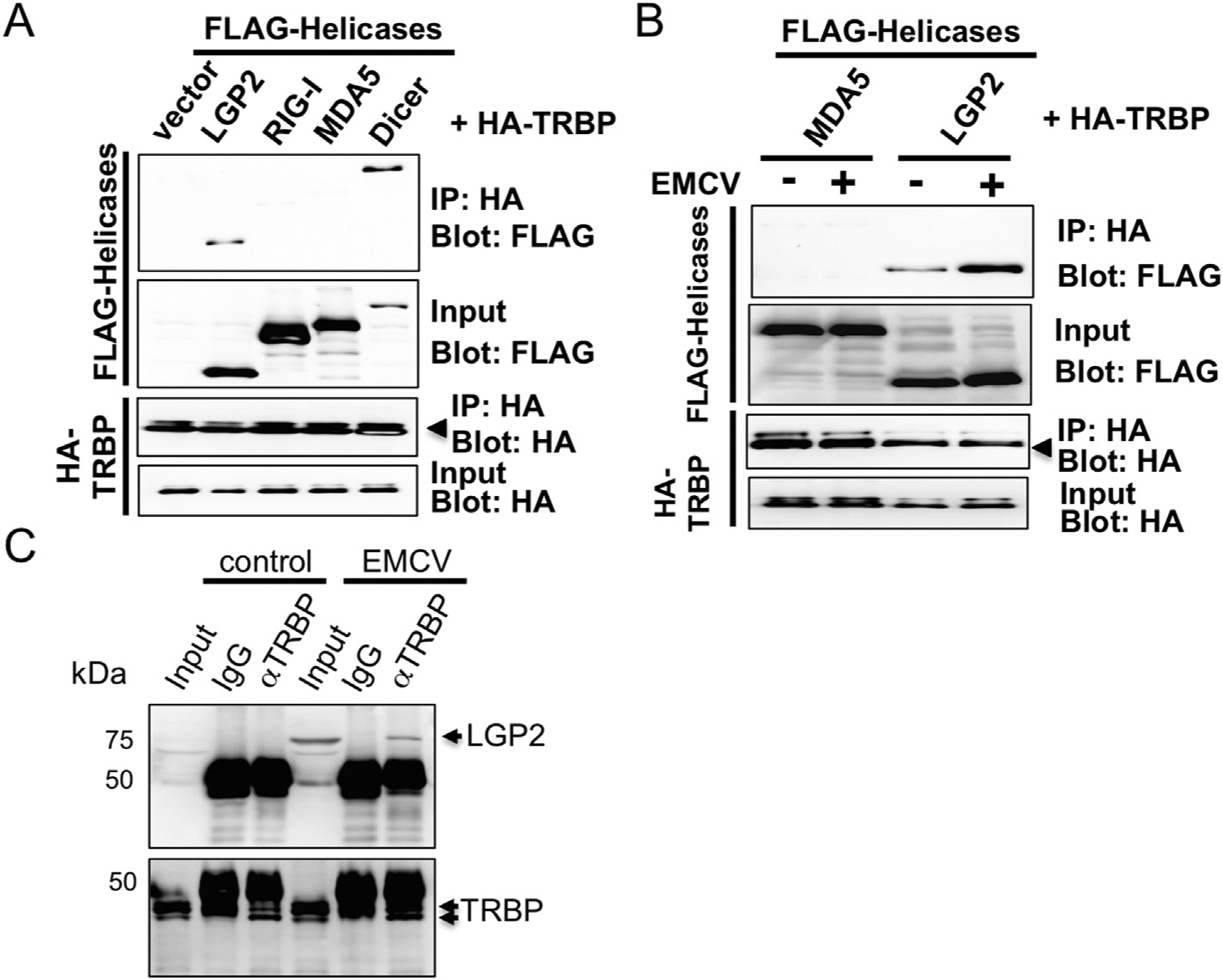
HA-tagged TRBP plasmid was co-introduced with FLAG-tagged LGP2, -RIG-I, MDA5 or Dicer plasmid in HEK293T cells and the lysate was subjected to immuno-precipitation with HA beads to co-purity FLAG-tagged proteins for Western blot (A). HA-tagged TRBP plasmid was co-introduced with FLAG-tagged LGP2 or MDA5 plasmid in HEK293T cells and cells were infected or mock infected with EMCV (MOI = 1) for 9.5 h. The lysate was subjected to immunoprecipitation as in A (B). The intensity was determined by densitometric analysis using Quantity One software (Bio-Rad). L929 cells were mock infected or infected with EMCV (MOI = 1) and the lysate was subjected to immunoprecipitation with anti-TRBP antibody or control rabbit IgG to co-purity endogenous LGP2 protein. The precipitant was probed with anti-LGP2 antibody for Western blot analysis (C).