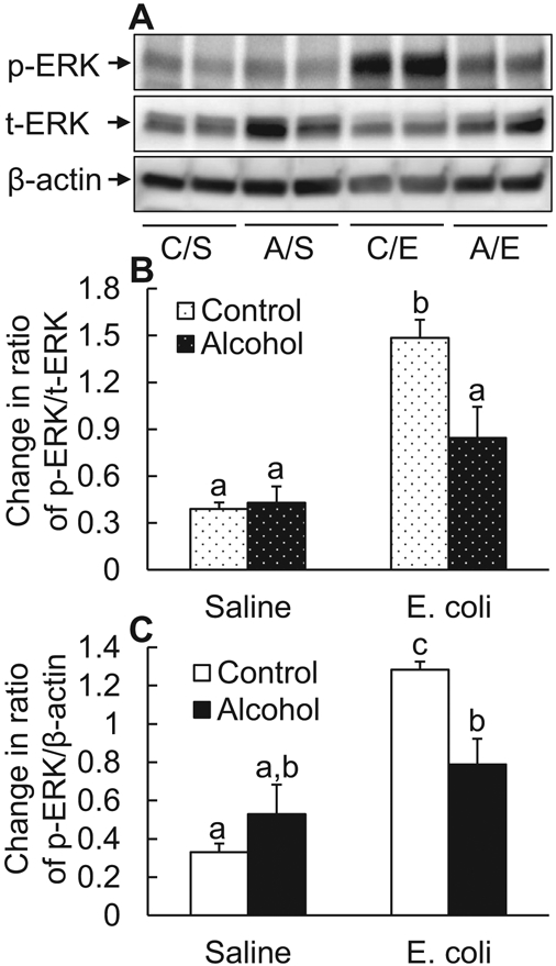
Fig. 2.

Acute alcohol intoxication suppressed ERK1/2 activation in nucleated bone marrow cells 8 hours following i.v. challenge with E. coli. (A) Representative Western blot image. C/S: i.p. saline plus i.v. saline; A/S: i.p. alcohol plus i.v. saline; C/E: i.p. saline plus i.v. E. coli; A/E: i.p. alcohol plus i.v. E. coli. (B and C) Quantitative analysis of Western blot images. Control: i.p. saline; alcohol: i.p. alcohol; saline: i.v. saline; E. coli: i.v. E. coli. Values are mean ± SEM. N = 4 in each group. Bars with different letters in each panel of (B) and (C) are statistically different (p < 0.05).
