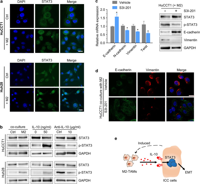
Fig. 7.
STAT3 signaling is critical for M2 macrophage-induced EMT in ICC cells. a IF staining for STAT3 in HuCCT1 and Huh28 cells cocultured with THP-1-derived M2 macrophages. The green color represents STAT3 staining, and the blue signal represents DAPI-stained nuclei. b Western blotting analyses of STAT3 and p-STAT3 expression in ICC cells cocultured with M2 macrophages treated with IL-10 and its neutralizing antibody. c IF staining for EMT markers in HuCCT1 cells cocultured with M2 macrophages after treatment with STAT3 inhibitor (S3I-201). d The left panel shows EMT marker expression in HuCCT1 cells after treatment with S3I-201, as determined by qRT-PCR, and the right panel shows the changes in p-STAT3 and EMT markers (E-cadherin and vimentin). e A schematic model describing the mechanism investigated in the present study in which ICC induces M2-polarized tumor-associated macrophages, and IL-10 secreted by M2 macrophages promotes tumor growth, invasiveness and EMT via STAT3 signaling. The data are presented as the mean ± SD; *p < 0.05. Bar = 1 mm

