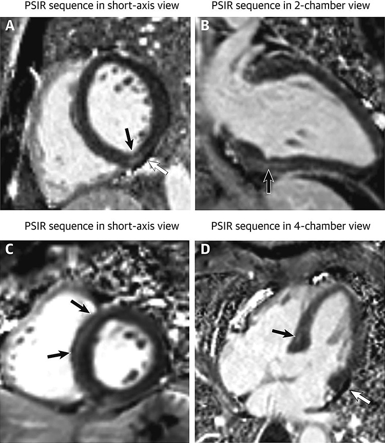
Figure 3.

Focal myocardial fibrosis in patients recovered from COVID-19: a 29-year-old male patient (first row) underwent cardiac CMR 1 month after the onset of palpitations. A 60-year-old male patient (second row) underwent cardiac CMR 2 months after the onset of palpitations. PSIR sequences in short-axis view (A, C) showed focal LGE (black arrows) in inferior and septal segments of left ventricle, respectively. Results were confirmed on the PSIR sequences in 2-chamber view (C) and 4-chamber view (D). Images A and D demonstrated a small pericardial effusion (white arrow) in both patients.; LGE, late gadolinium enhancement; CMR, cardiac magnetic resonance; PSIR, phase-sensitive inversion recovery. Image reproduced from Huang et al. [8] under the terms of the creative commons attribution 4.0 international license.
