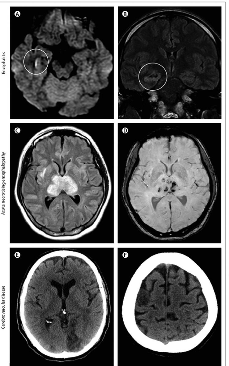
Figure 9.

Brain imaging in patients with neurological disease associated with COVID-19; (A) hyperintensity along the wall of inferior horn of right lateral ventricle on diffusion-weighted imaging, indicating ventriculitis; (B) hyperintense signal changes in the right mesial temporal lobe and hippocampus with slight hippocampal atrophy, consistent with encephalitis; (C) hyperintensity within the bilateral medial temporal lobes and thalami; (D) evidence of haemorrhage, indicated by hypointense signal on susceptibility-weighted images, consistent with acute necrotising encephalopathy; (E) CT showing ischaemic lesions involving the left occipital lobe; (F) right frontal precentral gyrus of the brain in a man aged 64 years who deteriorated neurologically after admission to hospital with COVID-19 and was diagnosed with acute stroke. Permissions granted to reproduce image from Ellul et al. [18]
