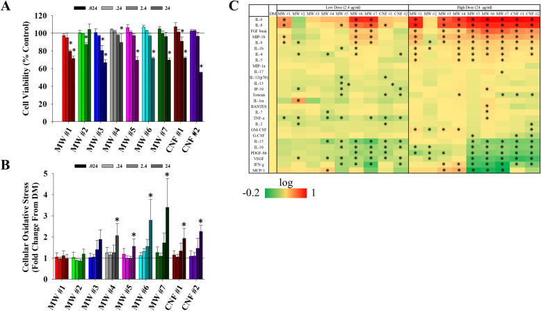
Fig. 7.
Toxicity assessment of BEAS-2B cells exposed to CNT/F. a WST-1 cell proliferation assay was used to assess the viability of BEAS-2B cells following exposure to increasing concentrations (0.024–24 μg/ml) of CNT/F. The dose at which the particle significantly reduced cell viability is indicated with an asterisk (p < 0.05). b Oxidative stress was measured using the CellROX assay. * p < 0.05 fold change vs. control cells represented as a reference line. c Protein secretions from cells exposed to 2.4 or 24 μg/ml of various CNT/F for 24 h represented as heat maps of fold change from controls with no exposure.. Significant changes from control cells were indicated with an asterisk (* p < 0.05). Log fold change was represented by color with green indicating a decrease in protein concentration and red indicating an increase on a scale of − 0.2 to 1. (*p < 0.05)

