
Fig. 4.
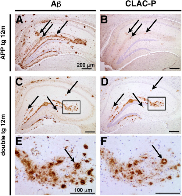
Endogenous and overproduced CLAC co-deposited with Aβ on the Aβ deposits. a–f Immunohistochemical analyses of the brains of 12-month-old APP tg (J20 line, a and b) and double tg mice (c–f) by anti-human Aβ (BAN50) (a, c and e) and anti-CLAC antibodies (anti-NC4) (b, d and f). High magnification images (insets in c and d) are shown in (e) and (f), respectively. In hippocampus of APP tg mice, immunoreactivities of murine endogenous CLAC were positive in a part of Aβ plaques (arrows in a and b). Middle-sized plaques in the hippocampus of double tg mice were visualized by anti-Aβ and anti-CLAC antibodies (arrows in c–f). Scale bar shows 200 μm (a–d) and 100 μm (e and f)