Fig. 5.
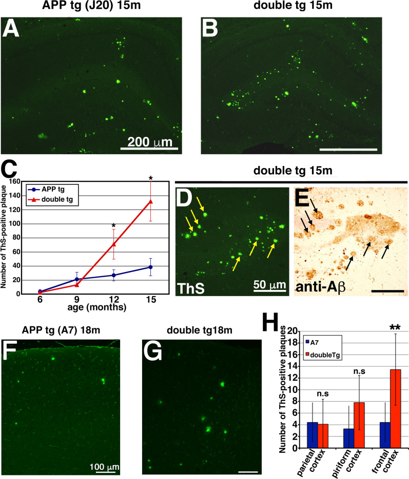
Overexpression of CLAC-P significantly increased the number of ThS-positive plaques. a, b ThS staining of the brains of 15-month-old APP tg (J20 line, a) and double tg mice (b). c The mean number of ThS-positive plaques was assessed in the hippocampus of 6-, 9-, 12- and 15-month-old APP tg mice (blue circles) and double tg mice (red triangles). N = 4, 4, 13, 7 for 6-, 9-, 12-, 15-month-old APP tg, respectively. N = 4, 3, 12, 7 for 6-, 9-, 12-, 15-month-old double tg, respectively. Student’s t-test, p = 0.81 (6-month-old), p = 0.24 (9-month-old), p < 0.0001 (12-month-old), p < 0.0001 (15-month-old). d, e Serial sections stained with ThS (d) and anti-Aβ antibody (BAN50) (e) were shown. The core-region of middle-sized plaques in the brains of 15-month-old double tg mice were exclusively labeled by ThS (arrows). f, g ThS staining of the brains of 18-month-old APP tg (f) and double tg (g) mice. h The mean number of ThS-positive plaques assessed in the parietal, piriform and frontal cortices of APP tg and double tg mice. N = 4, Student’s t-test, p = 0.86 (parietal cortex), 0.070 (piriform cortex), 0.00063 (frontal cortex), **, p < 0.01. Scale bar shows 200 μm (a and b), 50 μm (d and e), 100 μm (f and g)

