Fig. 6.
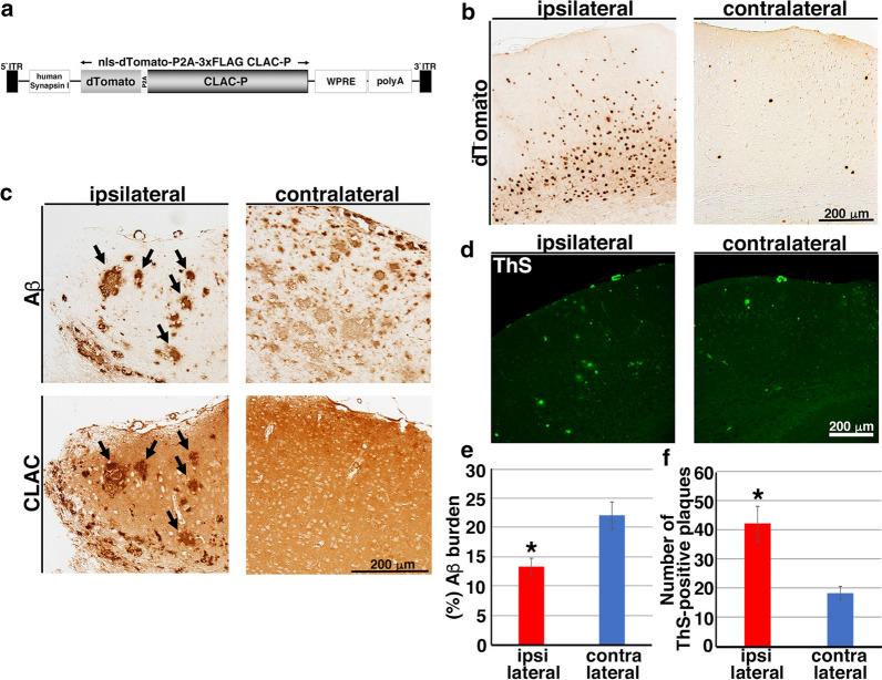
Overexpression of CLAC-P remodeled the morphology of pre-formed Aβ plaques. a A schematic structure of AAV9-CLAC-P. Under the human Synapsin I promoter, both dTomato and human CLAC-P are expressed bicistronically in neurons due to P2A self-cleaving peptide sequence. b Representative pictures of immunohistochemical staining of AAV9-CLAC-P-injected (ipsilateral) or PBS-injected (contralateral) hemisphere of 18-month-old APP tg mice (A7 line) using an anti-RFP antibody for dTomato. Scale bar shows 200 μm. c Immunohistochemical analyses of the brains of AAV9-CLAC-P-injected (ipsilateral) or PBS-injected (contralateral) 18-month-old APP tg mice (A7) using anti-Aβ (82E1, upper panels) or anti-CLAC antibodies (anti-NC3, lower panels). Arrows indicated the Aβ- and CLAC-double positive plaques. N = 5. Scale bar shows 200 μm. d ThS staining of the brains of AAV9-CLAC-P-injected (ipsilateral) or PBS-injected (contralateral) 18-month-old APP tg mice. Scale bar shows 200 μm. e Quantitative analysis of amyloid burden in the neocortex of AAV9-CLAC-P-injected (ipsilateral) or PBS-injected (contralateral) 18-month-old APP tg mice. N = 5, Paired t-test, p = 0.046, * p < 0.05. f The mean number of ThS-positive plaques in the neocortex of AAV9-CLAC-P-injected (ipsilateral) or PBS-injected (contralateral) 18-month-old APP tg mice. N = 5, Paired t-test, p = 0.017, * p < 0.05

