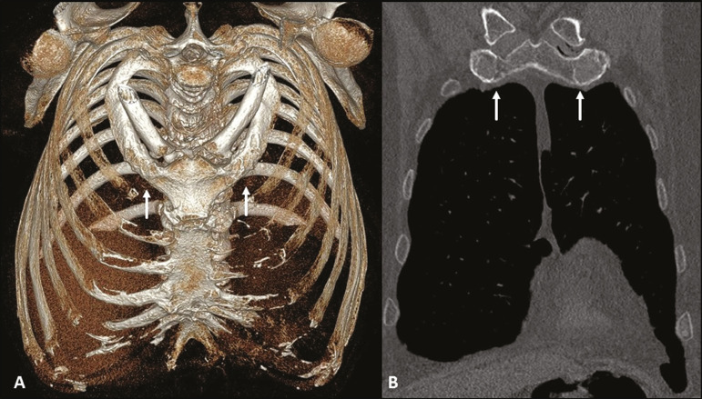
Figure 3.
Physiological costochondral calcifications. MDCT (A - three-dimensional reconstruction; B - coronal view) showing fusion of the first rib to the sternal manubrium (arrows) in an 83-year-old male patient, together with calcification of the other sternocostal and bilateral chondrocostal cartilages. Note the tramtrack pattern of calcification.

