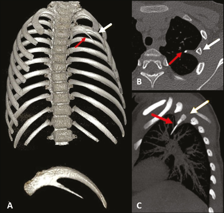
Figure 7.
Intrathoracic rib (type Ib). MDCT (A - three-dimensional reconstruction; B - axial view; C - sagittal maximum intensity projection) showing a supernumerary rib (red arrows) originating in the posterior portion of a short left third rib (white arrows), with a lateral, inferior oblique path to the pulmonary parenchyma. Note that the rib has no visible medullary cavity. No changes were identified in the posterior vertebral bodies or ribs.

