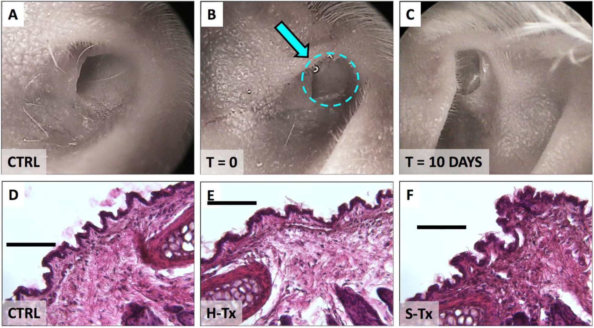
Figure 5. Physiological effects of thixogels of thixogel application into ears.

Top panels illustrate the otoscopic evaluation of the mouse ear canal: A – prior to thixogel application; B – immediately after thixogel application (thixogel highlighted by dotted area and indicated by the arrow); and C – 10 days post deployment. No detectable thixogel was present 10 days post-deployment. The bottom row illustrates the histological evaluation of tissue treated with D – saline (control); E – H-Tx and F – S-Tx. Scale bar is 100 μm.
