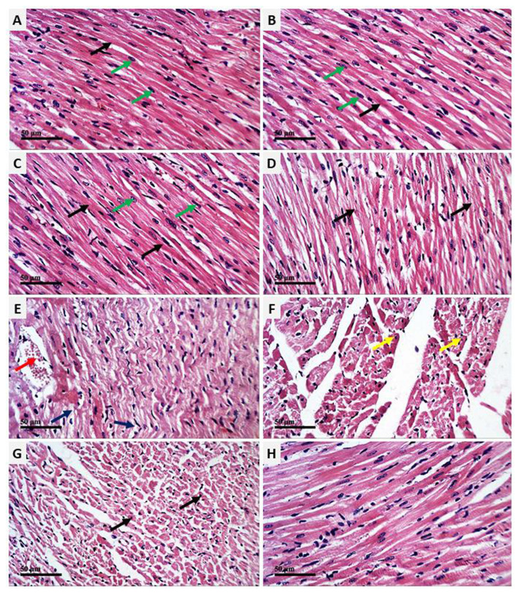
Figure 2.
AV and ED prevent CP-induced cardiac injury in rats. Photomicrographs of the heart tissue sections of control (A) and rats treated with AV (B) and ED (C) showing normal structure of the cardiomyocytes with normal nuclei (green arrow) and cytoplasmic striation (black arrow), CP-intoxicated rats (D–F) showing degenerative changes (blue arrow), fragmented myofibrils (black arrow), nuclear pyknosis (yellow arrow), increased intercellular spaces between adjacent cardiomyocytes and many congested intermuscular blood vessels (red arrow), and CP-intoxicated rats treated with AV (G) and ED (H) showing improved histological architecture of the heart with fewer degenerative changes and congestions (arrow). (H&E, Scale bar = 50 µm).

