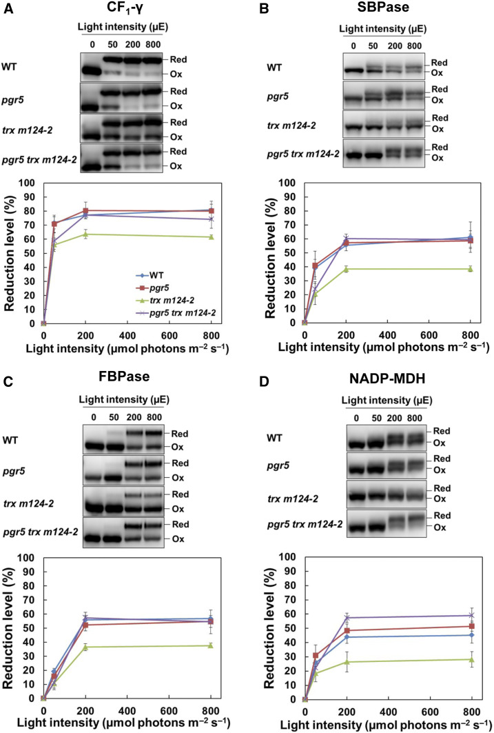
Figure 3.
Photo-Reduction of the Thiol Enzymes in the Wild Type and the pgr5, trx m124-2, and pgr5 trx m124-2 Mutants under Different Light Intensities.
(A) to (D) After 8 h in dark, seedlings were subjected to illuminations of 50, 200, and 800 µmol photons m–2 s–1 (µE), for 1 h each, in a stepwise manner. Samples were collected at the indicated light intensities and modified with AMS, after which 35 µg of proteins (per sample) were subjected to nonreducing SDS-PAGE. Redox states of the ATPC1 (A), SBPase (B), FBPase (C), and NADP-MDH (D) were detected by immunoblot analysis (top). The reduction levels of thiol enzymes were indicated as the percentage of the reduced versus the total protein (bottom). Each value represents the mean ± sd (n = 5 independent plants). Ox, oxidized; Red, reduced; WT, wild type.

