Figure 8.
Dynamics of Complex Formation between Trx m4 and PGRL1 during Photosynthesis In Vivo.
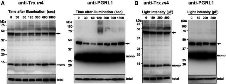
(A) Transient dissociation of the Trx m4-PGRL1 complex during the induction of photosynthesis. Seedlings of the wild type were dark adapted for 8 h and exposed to 200 µmol photons m–2 s–1 for the indicated time periods, before harvesting the plant materials. Next, 50 µg of protein samples was subjected to nonreducing SDS-PAGE. The Trx m4-PGRL1 complex was immunodetected using Trx m4 and PGRL1 antibodies. Arrows indicate the Trx m4-PGRL1 complex. The total amount of Trx m4 or PGRL1 protein (total) was analyzed on reducing SDS-PAGE to cleave disulfide bonds of complexes, in the same volume used in the detection of Trx m4-PGRL1 complex. At least three independent experiments using different plants were performed, and the representative results are shown.
(B) Trx m4-PGRL1 complex formation, under different light intensities. Seedlings were first dark adapted (8 h) and then illuminated at 50, 200, and 800 µmol photons m–2 s–1, for 1 h each, in a stepwise manner. Samples were collected at the indicated light intensities. Other details are as described in (A). To detect the Trx m4-PGRL1 complex, the chemiluminescence signal of PGRL1 monomer was oversaturated.

