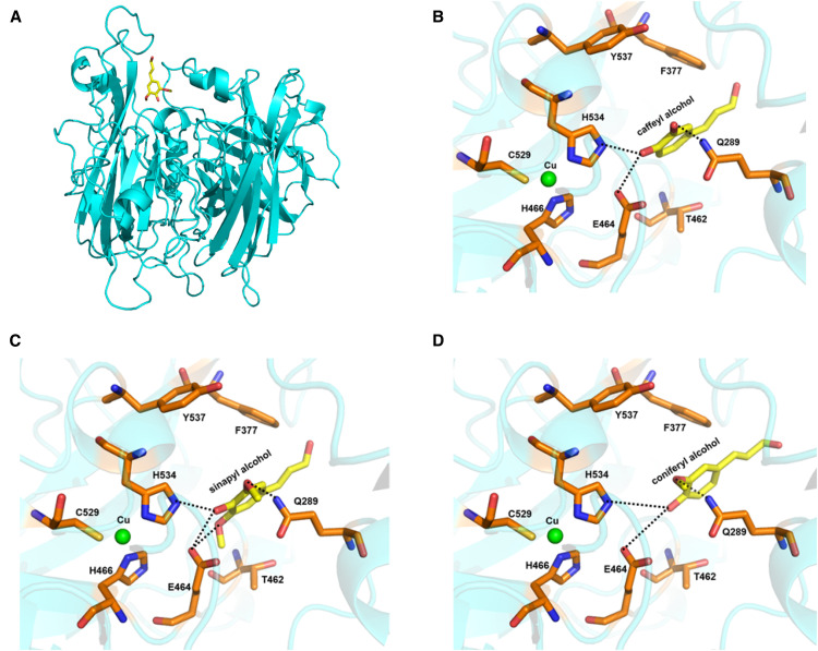
Figure 3.
Examination of the Binding of Monolignols in the Active Site of ChLAC8 by Molecular Modeling.
(A) A modeled structure of ChLAC8 with sinapyl alcohol docked into the active site.
(B) Active site residues showing binding of caffeyl alcohol.
(C) Active site residues showing binding of sinapyl alcohol.
(D) Active site residues showing binding of coniferyl alcohol.
Caffeyl alcohol, sinapyl alcohol, and coniferyl alcohol are shown as a stick model in yellow. Some key protein residues in the active site and binding pocket are labeled and shown as stick models in orange. T1 Cu is shown as a sphere model in green. The predicted hydrogen bonds are indicated by dashed lines.

