. 2020 Dec 1;9:e62048. doi: 10.7554/eLife.62048

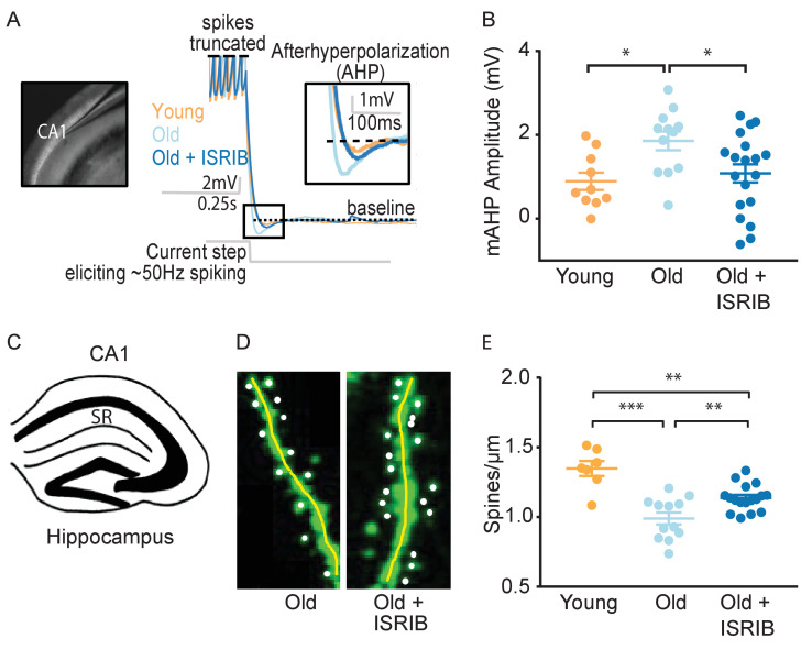
Figure 3. ISRIB treatment alleviates age-associated changes in CA1 pyramidal neuron function and structure.

(A) Left: Image of pipette patched onto CA1 neuron in sagittal slice of hippocampus. Right: Representative traces from hippocampal CA1 pyramidal neurons from old animals treated with either vehicle (light blue) or ISRIB (dark blue) or young animals treated with vehicle (orange) showing the response to a current injection eliciting ~50 Hz spiking activity. Spikes are truncated (dashed line), and the AHP is visualized immediately following cessation of current injection (yellow square) and quantified as the change in voltage from baseline (dotted line). (B) Age-induced increases in AHP were measured when comparing young and old animals. ISRIB treatment reversed increased AHP to levels indistinguishable from young animals. Animals were injected with ISRIB (2.5 mg/kg) or vehicle intraperitoneal 1 day prior to recordings. One-way ANOVA (F = 4.461, p<0.05); with Tukey post-hoc analysis. *p<0.05. Each neuron is represented with a symbol; lines indicate the mean ± SEM (Neurons: Young males n = 10 (5 animals); Old males n = 12 (5 animals), Old + ISRIB males n = 19 (7 animals)) with 1–5 neurons recorded per animal. (C–E) Spine density was quantified in the CA1 region of the dorsal hippocampus from young and old Thy1-YFP-H mice. (C) Diagram of hippocampal region analyzed. SR = stratum radiatum. (D) Representative images from Old and Old + ISRIB mice. (E) A decrease in dendritic spine density was measured when comparing old mice to young mice. ISRIB treatment significantly increased spine density levels of old mice when compared to vehicle-treated old mice. 63x magnification with a water immersion objective. Young males n = 7 slides (two animals); Old males + Vehicle n = 12 slides (three mice); Old males + ISRIB n = 17 slides (four mice). Individual slide scores (relative to old mice) represented in dots, lines depict group mean ± SEM. One-way ANOVA (F = 18.57, p<0.001) with Tukey post-hoc analysis. **p<0.01; ***p<0.001.

Figure 3—source data 1. List of electrophysiology reagents.
Column 1 – reagent name; Column 2 – Company of purchase; Column 3 – Catalog number.

Figure 3.

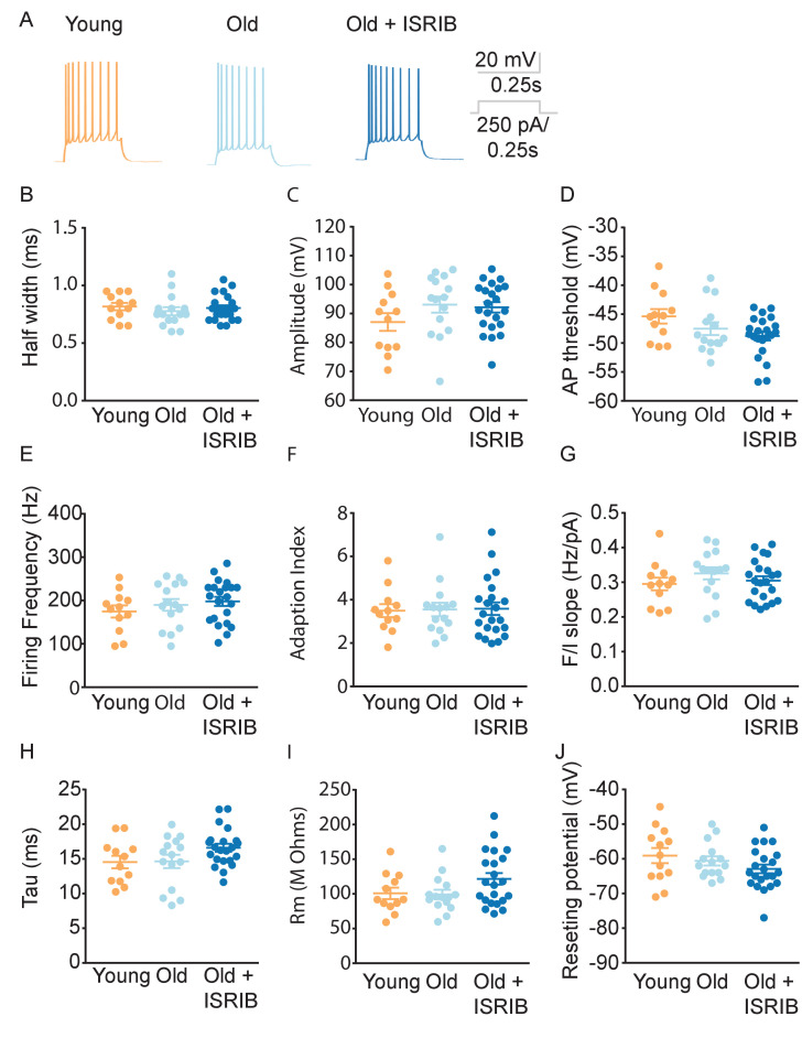
Figure 3—figure supplement 1. Age and ISRIB treatment do not modify other passive or active intrinsic membrane properties in CA1 pyramidal neurons.

Figure 3—figure supplement 1.

(A) Representative traces from CA1 pyramidal neurons showing the membrane potential response to a 250 pA current injection in neurons from old animals treated with either vehicle (light blue) or ISRIB (dark blue) or young animals treated with vehicle (orange). Quantification of the action potential (AP) including the half width (B), amplitude (C), and threshold (D) did not show significant differences between CA1 pyramidal recordings from old, old + ISRIB-treated, or young mice. Likewise, evaluation of the maximum firing frequency (E) or how the frequency of spiking changes over time, quantified by the adaptation index (F) or with current injection, quantified by the slope of the relationship of firing frequency versus amplitude of current injection (F/I slope) (G) was also not significantly different between groups. Finally, passive membrane properties including the membrane time constant (tau) (H), membrane resistance (Rm) (I), and the resting membrane potential (J) were not significantly altered by age or ISRIB treatment. Each neuron is represented with a symbol; solid lines indicate the mean ± SEM. (One-way ANOVA for all comparisons; Neurons: and Young males n = 12 (5 animals); Old males n = 15 (5 animals), Old + ISRIB males n = 22 (7 animals)) with 1–5 neurons recorded per animal.
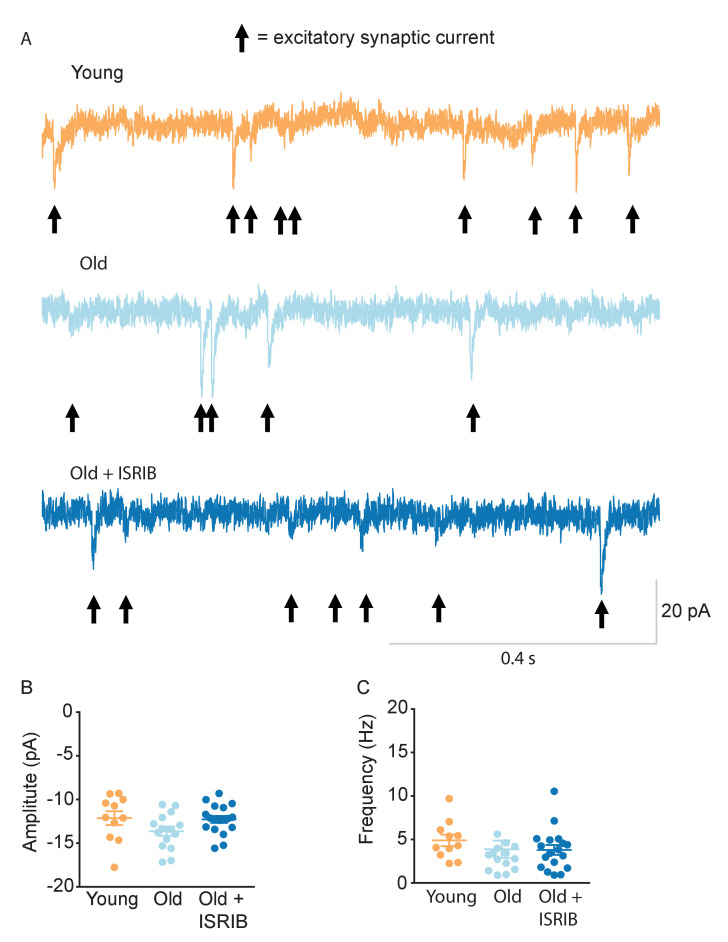
Figure 3—figure supplement 2. Age and ISRIB treatment do not affect spontaneous excitatory post-synaptic currents (sEPSC) in CA1 pyramidal neurons.

Figure 3—figure supplement 2.

(A) Representative whole cell voltage-clamp recordings showing sEPSCs from CA1 pyramidal neurons from old animals treated with either vehicle (light blue) or ISRIB (dark blue) or young animals treated with vehicle (orange). Arrows denote synaptic currents. (B) The sEPSC amplitude was not significantly different between groups (one-way ANOVA). (C) The sEPSC frequency was unchanged after ISRIB treatment or compared to young mice (Kruskal-Wallis test). The median amplitude or frequency for each neuron is represented with a symbol; solid lines indicate the mean ± SEM. (Neurons: Young males n = 11 (5 animals); Old males n = 15 (5 animals), Old + ISRIB males n = 18 (7 animals) with 1–5 neurons recorded per animal.).