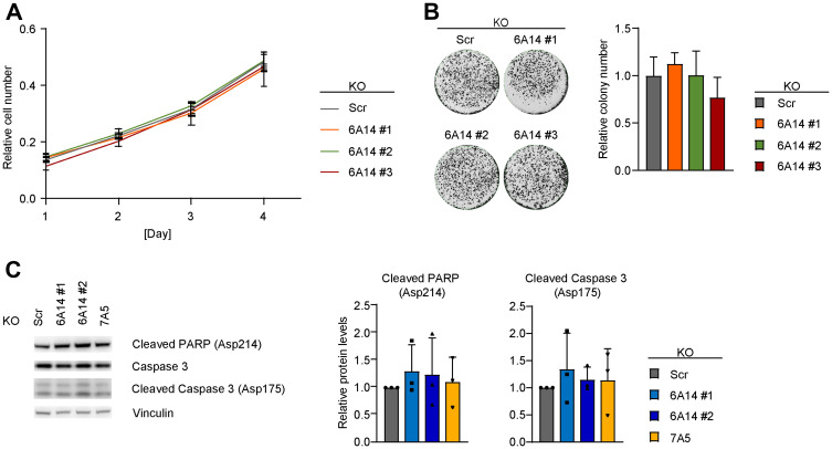
Figure 2. Genetic inhibition of SLC6A14 does not have a major impact on cell viability.
(A) Cell number quantification of scrambled and SLC6A14 KO MDA-MB-468 cell lines. Equal number of cells were plated, stained with crystal violet at each indicated time point and quantified by reading the absorbance at 550 nm. Data represent mean ± standard deviation of 3 independent experiments. (B) (Left) Representative colony formation assay image of scrambled and SLC6A14 KO MDA-MB-468 cell lines. (Right) Colony number quantification. Data represent mean + standard deviation of 4 independent experiments. (C) (Left) Immunoblot showing levels of apoptotic markers in inducible MDA-MB-468 KO cell lines after 5 days Dox-induction. (Right) Immunoblot quantification. Data represent mean + standard deviation of 3 independent experiments.

