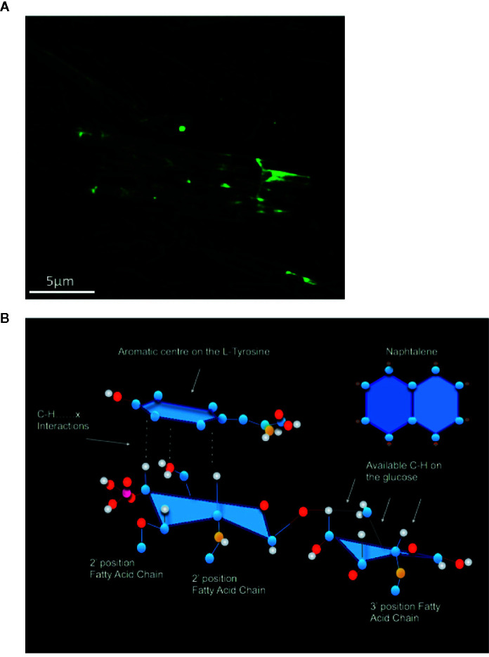
Figure 3.

(A) The physical association of MPL® across the needle-like crystalline structure of 20 mg/ml MCT® has been characterized using fluorescently labeled LPS (100 µg; Lipopolysaccharide) as a substitute for MPL® via confocal microscopy. (B) Proposed C–H⋯π interactions between the 2-deoxy-2-aminoglucose on MPL® and the aromatic ring on L-tyrosine, based on inhibitor studies with Naphthalene (Adapted from Bell et al., 2015).
