FIGURE 5.
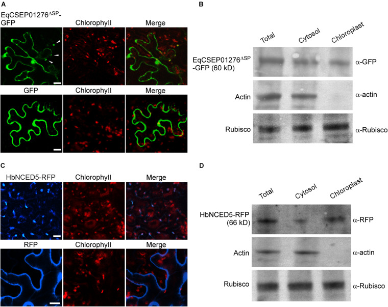
Localizations of EqCSEP01276 and HbNCED5 in plant cells. (A) Localizations of EqCSEP01276ΔSP-GFP and GFP in N. benthamiana leaves were examined by confocal microscopy. Each image of GFP or chlorophyll channel was photographed by using Z-stack tool of confocal fluorescence microscopy to scan one leaf area containing epidermal and mesophyll cells. White arrows indicate the chloroplast-localized EqCSEP01276ΔSP. Bars = 10 μm. (B) The total leaf proteins and proteins from cytosol and chloroplasts were extracted from N. benthamiana leaves expressing EqCSEP01276ΔSP-GFP. The resulting proteins were then analyzed by western blot using anti-GFP, anti-RFP, anti-β-actin, and anti-Rubisco antibodies. (C) Localizations of HbNCED5-RFP and RFP in N. benthamiana leaves were examined by confocal microscopy. Each image of RFP or chlorophyll channel was photographed by using Z-stack tool of confocal fluorescence microscopy to scan one leaf area containing epidermal and mesophyll cells. Bars = 10 μm. (D) The total leaf proteins and proteins from cytosol and chloroplasts were extracted from N. benthamiana leaves expressing HbNCED5-RFP. The resulting proteins were then analyzed by western blot using anti-GFP, anti-RFP, anti-β-actin, and anti-Rubisco antibodies.

