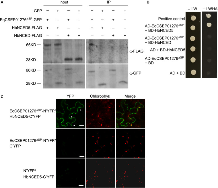
FIGURE 6.
The interaction between EqCSEP01276 and HbNCED5. (A) Co-immunoprecipitation for interaction between EqCSEP01276ΔSP and HbNCED5 or HbNCED. The total proteins from N. benthamiana leaves transiently expressing proteins of interest (Input) and the proteins eluted from anti-GFP beads were analyzed by western blot assay with anti-GFP or anti-FLAG antibodies. The GFP was used as the control. The sizes of protein bands: HbNCED5 (66 KD), HbNCED (28 KD), EqCSEP01276Δ SP-GFP (60 KD), GFP (28 KD). (B) In yeast two-hybrid assay, yeast transformants expressing EqCSEP01276ΔSP and HbNCED5 or HbNCED were assayed for growth on SD-LW or SD-LWHA. L, leucine; W, tryptophan; H, histidine; A, adenine. The combination of AD-T and BD-53 was used as a positive control, and the other sets were used as negative controls. (C) Bimolecular fluorescent complimentary (BiFC) assay of the interaction between EqCSEP01276ΔSP and HbNCED5. YFP signals were detected in the N. benthamiana leaves transiently co-expressing EqCSEP01276ΔSP-N’YFP and HbNCED5-C’YFP using confocal microscopy. The other sets were used as negative controls. Each image of YFP or chlorophyll channel was photographed by using Z-stack tool of confocal fluorescence microscopy to scan one leaf area containing epidermal and mesophyll cells. White arrows indicate the chloroplast-localized YFP. Bar = 10 μm.

