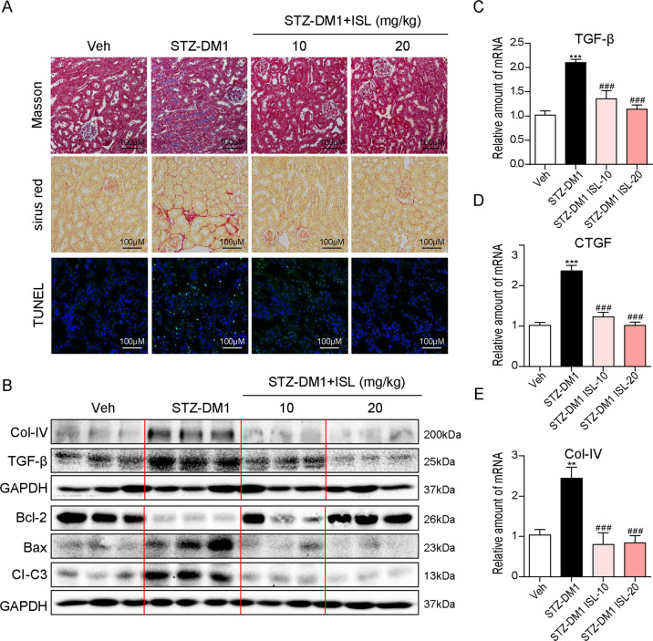
Fig. 2. ISL suppressed hyperglycemia-induced renal fibrosis and apoptosis.
a Representative images for Masson staining, Sirius red staining, and TUNEL staining using the formalin-fixed renal tissues as specified in Methods and materials (×200 magnification). b Western blot analysis for the protein expression of COL-IV, TGF-β, BCL-2, BAX, and CI-C3 in three kidney tissue extracts, each from a different mouse. c–e The mRNA expression of pro-fibrotic genes TGF-β, CTGF, and COL-IV in the kidney tissues was determined by real-time qPCR (A minimum of five mice per group were used for analysis. **P < 0.01, ***P < 0.001 vs control (Veh); ###P < 0.001 vs STZ1-DM).

