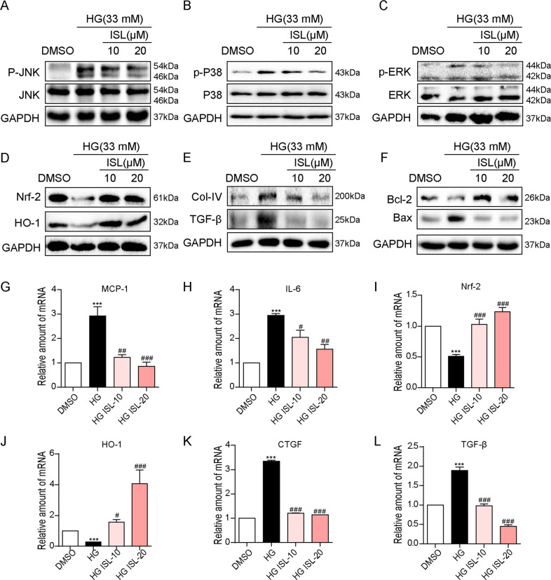
Fig. 4. ISL reduced HG-induced inflammation, oxidative stress, fibrosis, and apoptosis in NRK-52E cells.
NRK-52E cells (1 × 10⁶) were challenged with HG for the indicated time and subjected to 1 h pre-treatment with ISL (10 or 20 μM). a–c After 1 h HG-stimulated, western blot analysis was performed to investigate MAPK activation, showing the phosphorylation of p38 a, ERK b, and JNK c with the corresponding total protein as a loading control. d 12 h after HG-stimulated, western blot analysis was performed to study the expression of Nrf-2 and HO-1 with GAPDH as a loading control. e, f 36 h after HG-stimulated, western blot analysis was carried out to study the expression of pro-fibrosis genes, COL-IV and TGF-β, or apoptosis-related genes, BAX and BCL-2, with GAPDH as a loading control; g–l The mRNA levels of MCP-1 and IL-6 in 6 h after HG-stimulated g, h; or Nrf-2 and HO-1 in 4 h after HG-stimulated i, j; or CTGF and TGF-β in 12 h after HG-stimulated k, l were detected by RT-qPCR. Bars represented means ± SEMs of four independent experiments (***P < 0.001 vs DMSO group; #P < 0.05, ##P < 0.01, ###P < 0.001 vs HG group).

