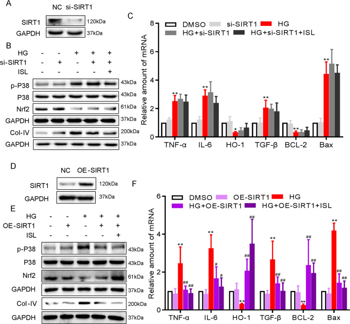
Fig. 6. ISL inhibited HG-induced inflammatory and oxidative injuries in NRK-52E cells via SIRT1.
NRK-52E cells (1 × 10⁶) were challenged with HG for the indicated time and subjected to 1 h pre-treatment with ISL (10 μM). a NRK-52E cells were transfected with siRNA against SIRT1. Control cells were transfected with negative control siRNA. Western blot was used to determine knockdown efficiency. b Immunoblot analysis of p-p38, p38, Nrf-2, and Col-IV following SIRT1 knockdown. c The mRNA expressions of TNF-α, IL-6, HO-1, TGF-β, Bcl-2, and Bax was determined by RT-qPCR at the same time as in Fig. 4. d NRK-52E cells were transfected with cDNA plasmids encoding SIRT1. Control cells were transfected with empty vector. Western blot was used to determine SIRT1 expression. e Immunoblot analysis of p-p38, p38, Nrf-2, and collagen IV following SIRT1 overexpression. f The mRNA expressions of TNF-α, IL-6, HO-1, TGF-β, Bcl-2, and Bax was determined by real-time qPCR at the same time as in c (*P < 0.05, **P < 0.01 vs DMSO group; #P < 0.05, ##P < 0.01 vs HG group).

